Journal of Pediatric Cardiology and Cardiac Surgery

Online ISSN: 2433-1783 Print ISSN: 2433-2720
Japanese Society of Pediatric Cardiology and Cardiac Surgery
Japanese Society of Pediatric Cardiology and Cardiac Surgery Academy Center, 358-5 Yamabuki-cho, Shinju-ku, Tokyo 162-0801, Japan
Journal of Pediatric Cardiology and Cardiac Surgery 1(1): 49-60 (2017)
doi:10.24509/jpccs.170110

ReviewReview

Preclinical Coronary Artery Anomalies and Silent Myocardial Ischemia in Children: How Can We Identify the Potentially Life-Threatening Conditions?

Nemours Cardiac Center, Nemours/Alfred I. duPont Hospital for Children ◇ USA

発行日:2017年7月3日Published: July 3, 2017
HTMLPDFEPUB3

Abnormalities of coronary artery, congenital or acquired, are rare in the pediatric population, but they frequently present unexpectedly. They may result in life-threatening conditions if unrecognized; however, in infants and children, preclinical coronary abnormalities usually do not present with the typical clinical manifestations seen in adults with myocardial ischemia. Timely diagnosis of silent myocardial ischemia is frequently challenging in children. Myocardial infarction may occur in anomalous left coronary artery from pulmonary artery (ALCAPA), right ventricle-dependent coronary artery seen in pulmonary atresia with intact ventricular septum (PA/IVS), and coronary aneurysm complicated in Kawasaki disease. On the other hand, some coronary artery abnormalities tend to present with unexpected ventricular arrhythmia or sudden cardiac death (SCD). These include aberrant aortic origin of coronary artery (AAOCA) and acquired coronary stenosis after arterial switch operation (ASO) for transposition of the great arteries (TGA), and post-transplant cardiac allograft vasculopathy (CAV), in which affected patients develop cardiac arrest or SCD without anginal pain or before having myocardial infarction. The pathological outcomes of myocardial ischemia in children are significantly different from those in adults with an atherosclerotic coronary disease; thus, the early recognition and detection of preclinical coronary anomalies and silent myocardial ischemia are essential. The underlying mechanism that determines myocardial responses to tissue ischemia involves complex processes, but it is plausible that mitochondrial signaling plays a critical role in determining these pathological paths. The involvement of the autonomic nervous system and the degree of collateral vessel development are contributing factors. In this review, pathobiology of myocardial ischemia and subsequent events will be discussed. The current paradigm of “myocardial ischemia” needs to be revisited in the field of pediatric cardiology.

Key words: ischemia; coronary; myocardial Infarction; ventricular fibrillation (VF); sudden cardiac death (SCD); mitochondria

Introduction

Myocardial ischemia is rare in children, but its timely diagnosis is essential, as delayed diagnosis may result in a catastrophic outcome. In adults, ischemic heart disease is the most frequent cause of morbidity and mortality in developed countries. Ischemic heart disease in adults is commonly due to coronary atherosclerosis and may present with recurrent chest pain (angina pectoris) or myocardial infarction as acute events, or with a chronic outcome including ischemic cardiomyopathy and congestive heart failure. Unexpected sudden cardiac death (SCD) is reported in some adult cases of coronary artery disease, especially during exercise.1, 2) In children, coronary atherosclerosis is very rare. Coronary anomalies in children can be classified as either congenital or acquired, and its clinical manifestation is diverse and quite different from that in adults.3, 4) Some coronary anomalies are diagnosed unexpectedly after life-threatening events or sudden death, whereas others present with asymptomatic heart murmur. Although rare, it is imperative for all practitioners to recognize the specific features of coronary anomalies in children; however, a reliable screening strategy for these disorders is currently not available.

In this review article, various coronary anomalies in children are introduced. Then, the underlying cellular pathobiology of myocardial ischemia and its clinical outcome with specific contributing factors are discussed. As an underlying pathological process, acute myocardial ischemia can introduce an ischemic tissue injury resulting in myocardial infarction as well as an electrical instability causing ventricular fibrillation and/or SCD. Lastly, the critical role of mitochondria in modulating pathological pathways in response to myocardial ischemic injury is outlined.

Overview of Coronary Abnormalities in Childhood

Although rare, there are a variety of coronary anomalies encountered during childhood. These can be either congenital or acquired. Several review articles are published on this topic.3, 5–7) In the following, certain representative coronary anomalies seen in childhood with specific clinical outcomes are illustrated.

Congenital Coronary Anomalies

Anomalous Left Coronary Artery from Pulmonary Artery

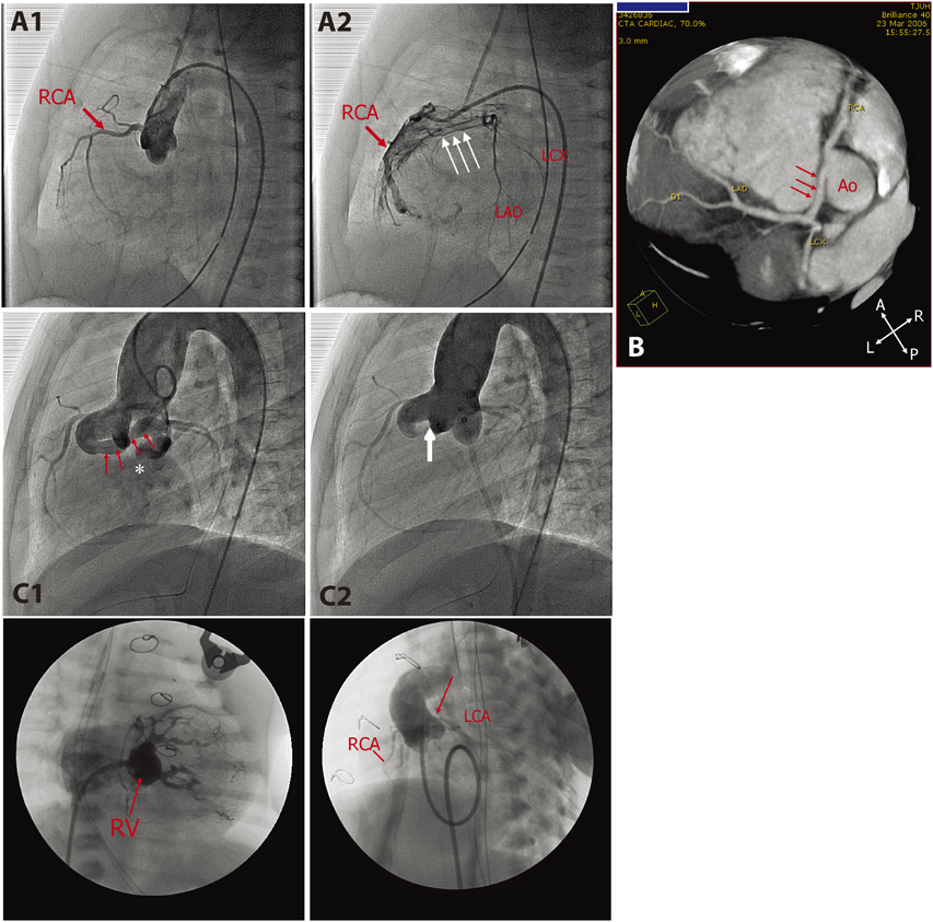
Anomalous left coronary artery from pulmonary artery (ALCAPA) is a rare congenital coronary anomaly typically presenting in infants around 2 to 3 months of age with acute onset of respiratory distress, poor feeding, and paleness secondary to circulatory failure.8–10) These clinical manifestations are attributed to myocardial infarction due to direct myocardial ischemic injury and associated left ventricular (LV) dysfunction and mitral regurgitation (MR). Early diagnosis and prompt surgical intervention (coronary translocation surgery) are essential for the better prognosis (Fig. 1A1, A2). Some infants completely recover LV function after an early diagnosis and a prompt surgical intervention.11–14) It is surprising that infantile myocardium regenerate, even after extensive myocardial damage.15)

Journal of Pediatric Cardiology and Cardiac Surgery 1(1): 49-60 (2017)

Fig. 1 Congenital coronary anomalies in children. A1 and A2: Coronary angiograms of 3-month-old male infant with anomalous left coronary artery from pulmonary artery (ALCAPA) who presented with irritability, respiratory distress, paleness, and feeding difficulty. Aortic root injection revealed only right coronary artery (RCA) arising from aorta (A1). Selective right coronary angiogram filled left anterior descending artery (LAD) and left circumflex artery (LCX) retrograde via collateral vessels (arrows). B: CT coronary angiogram of 19 years old asymptomatic male with anomalous aortic origin of coronary artery (AAOCA). Left coronary artery arises from the right coronary cusp and runs between the great vessels (arrows). C1 and C2: Aortic root injection before (C1) and after (C2) the deployment of a vascular occlusion device (a white arrow in C2) in the dilated coronary arterial fistula (CAF; arrows) from RCA to right atrium (*) in asymptomatic 3 years old boy who was diagnosed at 2 months of age because of continuous murmur. D1 and D2: A 4-day-old full term male infant with pulmonary atresia with intact ventricular septum and RV-dependent coronary circulation (RVDCC). D1: RV injection shows sinusoidal vessels communicating to native coronary artery system. D2: Aortic root injection reveals mild proximal narrowing of left coronary artery (LCA: arrow), suggestive of RVDCC

Patients with ALCAPA can be totally asymptomatic during childhood. There may be a mild cardiomegaly secondly to the increased left to right shunt with no clinical signs of myocardial ischemia or congestive heart failure. These patients are often encountered in adulthood with difficulty breathing, exercise intolerance, palpitation, chest pain, syncope,16–18) or even SCD.19) Associated late mitral valve insufficiency may complicate a clinical picture of chronic ischemic cardiomyopathy after initial surgical repair in early childhood.12, 20) Diversity of clinical presentation of ALCAPA is thought to depend on the degree of collateral vessel development, the extent of irreversible myocardial ischemic injury and fibrosis, associated hemodynamic abnormalities (pulmonary hypertension,21) large ventricular septal defect,22) patent ductus arteriosus,23) or severe branch pulmonary stenosis24)), and increased myocardial oxygen demand from LV dilatation. ALCAPA includes a clinical spectrum of significant spatiotemporal heterogeneity of myocardial ischemia due to congenital coronary anomaly.

Aberrant Aortic Origin of Coronary Artery

Aberrant aortic origin of coronary artery (AAOCA), another rare congenital coronary anomaly, occurs when the coronary artery arises from the different aortic cusps, consisting of left coronary artery (LCA) from the right aortic cusp and right coronary artery (RCA) from the left aortic cusp. This anomaly is frequently associated with other anatomical abnormalities including interarterial course, intramural course, a slit-like coronary orifice, and an acute angle from the aorta, which are speculated to be the underlying mechanisms of sudden disruption of coronary blood flow at the peak of exercise.25–27) The majority of patients with this condition remain asymptomatic, but the first symptom may be cardiac arrest or SCD, particularly aberrant aortic origin of left coronary artery (AAOLCA)28, 29) (Fig. 1B). AAOLCA is the second most common disease responsible for otherwise healthy young athletes during exercise (first is hypertrophic cardiomyopathy).30) The affected patients seldom experience ischemic chest pain or other warning signs of myocardial ischemia before the fatal event. AAOLCA is generally indicated for surgical intervention (coronary translocation or unroofing of intramural artery) because of a high association with SCD in otherwise healthy individuals.25) The surgical indication for aberrant aortic origin of right coronary artery (AAORCA) remains controversial.31)

The ultimate challenge of AAOLCA is difficulty in identifying this condition before a lethal event, as there are virtually no associated clinical signs or symptoms or laboratory biomarkers other than abnormal coronary imaging studies. The first clinical presentation may be an unexpected SCD. A common encounter of AAOLCA by pediatric cardiologists is either as a coincidental echocardiographic finding for unrelated problems (heart murmur, musculoskeletal chest pain, or nonspecific palpitations, etc.) or as a pathological finding at autopsy for the sudden death victims. Thus far, there is no effective screening method to detect this congenital anomaly.

Coronary Artery Fistula

Coronary artery fistula (CAF) or coronary cameral fistula implies fistulous communication between the existing coronary artery to any of the cardiac chambers (right atrium, left atrium, right ventricle, and left ventricle) or vessels (pulmonary artery)32, 33) (Fig. 1C1, C2). It can be small or enlarged with aneurismal morphology. Usually, CAF is diagnosed in asymptomatic patients with continuous murmur during childhood or clinical signs of left ventricular volume overload in adult patients. Adult patients may present with exercise intolerance or atrial arrhythmia due to volume overload and/or myocardial ischemia secondary to myocardial steal phenomenon.33) Echocardiography with a color Doppler study is usually sufficient to make a diagnosis. In children with asymptomatic CAF, fistula may be electively occluded with closure-device or coils under cardiac catheterization or surgically ligated. Symptomatic adult patients require necessary intervention. An understanding of correct coronary anatomy is crucial for intervention, as postoperative thromboembolic complications are reported.34–36)

Pulmonary Atresia with Intact Ventricular Septum

Variable coronary abnormalities are known to complicate in pulmonary atresia with intact ventricular septum (PA/IVS). Persistent exposure to significantly elevated right ventricular pressure predisposes the development of sinusoidal communication between the right ventricular cavity and existing coronary arteries (Fig. 1D1, D2).37) When this sinusoidal communication becomes predominant, the intrinsic coronary arteries may develop regression, causing proximal coronary stenosis or coronary atresia.38, 39) This condition is called “right ventricle-dependent coronary circulation (RVDCC),” and is responsible for an increased risk of myocardial ischemia or steal phenomenon with RV-decompression surgery or even shunt placement.40–42) This underlying coronary anomaly is responsible for high perioperative morbidity and mortality. It is essential to assess the possibility of RVDCC prior to surgical intervention to minimize the risk of myocardial infarction during and after surgery.43)

Acquired Coronary Anomalies

Acquired conditions that induce coronary abnormalities during childhood include Kawasaki disease and other vasculitis (Takayasu disease44)), coronary artery obstruction after arterial switch operation for transposition of the great arteries, post-transplant allograft vasculopathy, and genetic disorders (William syndrome45)).46) Atherosclerotic coronary disease is rare in children, although it may be seen in adolescents with familial hypercholesterolemia (homozygous type).47)

Kawasaki Disease and Giant Coronary Aneurysm

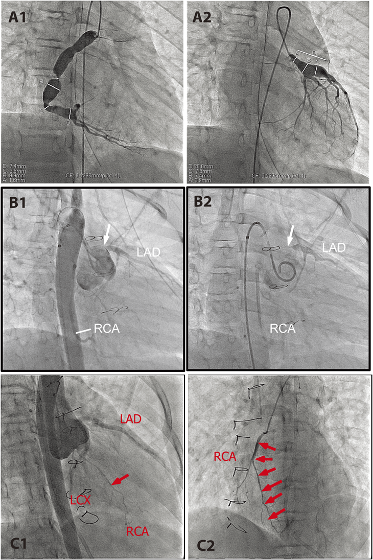
Kawasaki disease is known to cause coronary artery deformation, ectasia, and/or aneurismal formation during and after the acute phase of the disease.48, 49) Coronary aneurysm and secondary coronary stenosis as the results of uneven regression of ectasia are thought to predispose to vascular occlusion (Fig. 2A1, A2). Aneurysms are mainly seen at the proximal coronary arteries or at the bifurcations. Echocardiogram is useful in detecting these coronary lesions that evolve dynamically over time.50) Aneurysm may increase in size over the first 2 months, depending upon the degree of vasculitis, followed by spontaneous regression by 2 years after the onset; however, vascular stenosis progresses over many years.50, 51) Due to the early introduction of high-dose intravenous immunoglobulin (IVIG), the incidence of coronary aneurysm and its complications are dramatically decreased.52, 53) Vascular occlusion by thrombus formation and/or intimal thickening results in myocardial ischemia, infarction, or sudden cardiac death.54) Of 60 patients who developed myocardial infarction following Kawasaki disease, 50% developed within 6 months after the onset. Fifteen patients (25%) died, 3 died during the acute phase, and 12 patients died during the chronic phase. Half of the late deaths (6 patients) were of sudden unexpected death.54) The late sudden cardiac death frequently occurs in asymptomatic patients.55)

Journal of Pediatric Cardiology and Cardiac Surgery 1(1): 49-60 (2017)

Fig. 2 Acquired coronary anomalies in children. A1 and A2: Selective coronary angiograms of a 9-year-old girl with Kawasaki disease who developed a giant aneurysm during the acute phase (A1: RCA and A2: LCA). B1 and B2: Aortic root injection an 8-year-old girl with d-TGA who underwent aortic switch operation in the newborn period who developed sudden cardiac arrest during exercise. She had been totally asymptomatic until this near-lethal event. Filling of LCA was delayed by a few seconds with severe ostial stenosis. No collateral vessel formation was noted. C1 and C2: Coronary angiograms of a 12-year-old girl who underwent orthotopic heart transplant for dilated cardiomyopathy at 10 years of age. C1: Aortic root injection showed normal-sized LCA and diffusely hypoplastic RCA (C1), which was confirmed by selective RCA angiogram (C2)

d-Transposition of the Great Arteries after Arterial Switch Operation

Arterial switch operation (ASO) is primarily indicated in the newborn period for surgical repair of d-loop transposition of the great arteries (d-TGA) and Taussig-Bing anomaly. Because the procedure involves translocation of the coronary arteries, coronary complications may occur during the acute56, 57) and chronic phases.58–60) While short-term coronary complications are related to intrinsic coronary artery anatomy and surgical procedure per se, long-term complications frequently occur unexpectedly without any associated clinical signs or symptoms58, 61, 62) (Fig. 2B1, B2). Routine ECG, echocardiogram, exercise stress test (EST), and myocardial perfusion imaging (MPI) are neither sensitive nor specific to identify most of preclinical coronary disease.58, 63) Affected patients are usually asymptomatic or unexpectedly present with lethal (sudden death) or near-lethal (ventricular fibrillation) events. Pathological specimens reveal an intimal thickening of obliterated coronary arteries,60, 64) but the underlying mechanism of this process remains largely unknown. The necessity of routine coronary imaging study in post-ASO patients is advocated by certain groups, including the current author, to risk-stratify a preclinical coronary disease after ASO.60, 65, 66)

Cardiac Allograft Vasculopathy

Cardiac allograft vasculopathy (CAV), a diffuse coronary narrowing known to occur in the recipients of heart transplant, is a major risk factor in limiting long-term survival after heart transplantation.67–69) Unlike atherosclerotic coronary disease, CAV causes diffuse narrowing of coronary arteries (Fig. 2C). Histopathologically, CAV is characterized by the proliferation of vascular smooth muscle cells in the allograft and concentric intimal thickening.70) The pathogenesis of diffuse intimal thickening associated is thought to be due to multiple factors including chronic rejection, endothelial dysfunction, infection, and traditional cardiac risk factors (hypertension, hypercholesterolemia, and diabetes mellitus),67, 71) but the precise mechanism is not fully understood. The incidence of CAV in children is less than that in adults, but is known to occur in 5% to 7% within 2 years after transplant and 15% to 18% within 5 years after transplant.72) The prognosis of CAV is poor, as approximately 50% of cases will result in either graft loss or death within 2 years after the diagnosis.72, 73) Clinical presentation is variable and frequently nonspecific, as the affected patients may not complain of typical anginal pain because renervation over the implanted heart is often incomplete.74) The presence of abdominal, chest, and/or arm pain after heart transplant, however, is strongly associated with CAV and sudden death or aborted sudden death.75) The effective treatment or prevention for CAV is currently not known, but new data are emerging that can potentially help control or attenuate the development of pathological vascular remodeling.76)

Clinical Outcome and Underlying Mechanism of Myocardial Ischemia

Clinical manifestations of coronary disease in children are diverse and much different from those in adults. Although rare in incidence, failure to recognize the condition may result in an unexpected catastrophic outcome. Most children with myocardial ischemia do not necessarily complain of ischemic chest pain as a preceding warning symptom. They tend to be asymptomatic until they present abruptly with ventricular fibrillation or unexpected sudden death as a first presentation. SCD in middle-aged athletes presents in a similar way, as they are commonly considered least at risk to develop coronary insufficiency.1) There are two major challenges in managing silent coronary anomalies and preclinical myocardial ischemia in children: 1) difficulty in identifying the population at risk for SCD, and 2) lack of general guidelines in making risk stratification for SCD in patients with silent coronary anomalies.

Clinical Presentation of Myocardial Ischemia in Children

Heterogeneity of the ischemic myocardium and variable different physical conditions may create a diverse clinical spectrum encompassing angina chest pain, ventricular arrhythmia, myocardial infarction, cardiogenic shock, and SCD. SCD due to myocardial ischemia is seen with or without associated myocardial infarction, but usually occurs before the actual myocardial damage becomes evident.77) In other words, upon acute disruption of myocardial perfusion, some patients tolerate initial risk of developing ventricular fibrillation to survive until myocardial infarction develops.77)

Coronary anomalies in children may be classified into two groups based on the outcome of myocardial ischemia. The first, ALCAPA (early infancy), RVDCC in PA/IVS (early infancy), and giant aneurysm in Kawasaki disease (acute and chronic phase), may primarily present with myocardial infarction. The second group, AAOLCA, d-TGA after ASO (long term complication), and CAV, frequently present with unexpected cardiac arrest or SCD without visible myocardial infarction.78) Two cases of unexpected SCD were reported in Kawasaki disease with CA obstruction in a chronic phase.55) Upon myocardial ischemia due to sudden disruption of coronary blood flow, under what mechanisms can these two distinct pathways take place? It is imperative to understand the predisposing factors that favor ischemic myocardium developing into VF before having tissue injury.

Pathobiology of Myocardial Ischemia

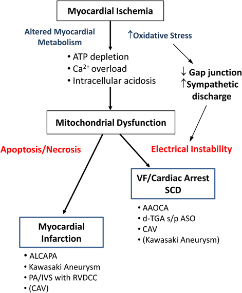
The heart is a highly energy-demanding organ that supports continuous dynamic pumping activity, in which oxygen delivery via coronary circulation plays a vital role. Sudden disruption of coronary blood flow causes myocardial ischemia, resulting in a mismatch between supply and demand of oxygen. Persistent disruption of myocardial perfusion not only induces energy depletion in each myocyte but also compromises washout of metabolic substrates generated by an anaerobic metabolism. As a result, ischemic myocytes develop intracellular acidosis, calcium overload, loss of gap junction function, irreversible cellular injury, increased oxidative stress, accumulation of reactive oxygen species (ROS), and mitochondrial dysfunction.79) These initial myocyte changes are responsible for two major pathological pathways: an electrical instability causing ventricular fibrillation (VF) and actual myocardial damage by cell death program (apoptosis and necrosis) (Fig. 3). Reperfusion following ischemia is primarily beneficial to reverse myocytes at risk, but it can be detrimental by introducing enhanced inflammatory responses inducing additional myocardial damage (ischemic-reperfusion injury).80) VF is an ominous condition, frequently responsible for SCD following myocardial ischemia.

Journal of Pediatric Cardiology and Cardiac Surgery 1(1): 49-60 (2017)

Fig. 3 Myocardial ischemia-induced electrical instability (VF and SCD) and direct myocardial tissue injury via apoptosis and necrosis. Mitochondria play a critical role in both pathological pathways

Several other factors may affect the fate of myocardium upon sudden disruption of coronary perfusion, including preconditioning, autonomic nervous system involvement, degree of collateral vessel development, and associated physical condition (e.g., exercise, emotional stress, cold weather). Preconditioning, a repetitive exposure of mild ischemic stress and reperfusion to enhance myocardial adaptation, is known to protect against greater subsequent ischemic insults with less threat of infarction81) and VF.82) Increased sympathetic discharge in association with pain, fear, and emotional outrage by sudden ischemia increases a risk of ventricular fibrillation or progression of direct myocardial injury.83) In an experimental myocardial infarction by left coronary artery ligation in the rat model, the incidence of sudden death was significantly higher under conscious sedation than under general anesthesia, suggesting detrimental effects induced by pain and associated sympathetic discharge.84) In ischemia-induced VF, sympathetic activation is considered proarrhythmic, whereas parasympathetic activation is antiarrhythmic.85) Targeted ablation of cardiac sympathetic neurons significantly reduced the susceptibility to ischemia-induced ventricular tachycardia in conscious rats.86) Active involvement of the autonomic nervous system in various cardiovascular diseases, including myocardial ischemia-induced SCD, was recently reviewed by Fukuda et al.87) Another important factor is collateral vessel formation or arteriogenesis, which is based on growth and remodeling of preexisting collateral anastomoses.88) The degree of collateral vessel development determines the myocardial integrity by increasing the threshold of electrical instability and cardioprotection against direct ischemic injury.89, 90) Preconditioning certainly enhances collateral vessel development, but the precise mechanism of arteriogenesis by ischemia is not fully understood.91) The overall net myocardial perfusion through obstructed native coronary arteries and the presence of collateral vessels may determine the final clinical outcome upon major ischemic events. Chronic myocardial ischemia commonly seen in ALCAPA, Kawasaki disease, or PA/IVS with RVDCC tends to stimulate collateral vessel formation, whereas AAOCA does not. The presence of collateral vessels increases cardioprotection against SCD,92) which has also been shown in CAV.93) Lastly, physical activities trigger SCD in coronary artery disease in older adults; this is more common in males and during heavier exercise.94) On the contrary, Reddy et al. reported that 80% of SCD occurred with light physical activities or during sleep rather than moderate to heavy exercise in older patients.95) The effects of exercise on SCD candidates are complex, as light to moderate regular exercise is proven to enhance cardioprotection against ischemic heart disease.96)

Mitochondria, a Critical Site of Regulating Myocyte Homeostasis

The Mitochondrion is an energy-converting machinery of cells that regulates metabolism via the Krebs cycle and electron-transport chains. Continuous vigorous myocyte contractile activity is highly dependent upon ATP generated by oxidative phosphorylation in the mitochondria with constant oxygen supply. In addtion to oxidative phosphorylation and ATP synthesis, mitochondria play a critical role in the regulation of Ca2+ and reactive oxygen species (ROS)-mediated processes in the myocytes.97, 98) Myocardial ischemia imposes multiple biological insults on mitochondria. As a result, hearts develop electrical instability responsible for VF and/or direct tissue injury through apoptosis and necrosis (oncosis) to induce myocardial infarction.99) Myocardial mitochondria play a vital role in determining which critical pathway to take in response to overwhelming ischemia. How can the energetic status of the mitochondrial network induce the myocardial electrical instability to cause VF and/or cell death signaling pathways to result in myocardial infarction?

Ischemia-induced oxidative stress alters the cardiac mitochondrial network and thus induces spatiotemporal action potential heterogeneity. This action potential heterogeneity causes lethal ventricular arrhythmia via multiple different levels including sarcolemmal ATP-sensitive potassium channel (sarcKATP), mitochondrial inner membrane anion channel (iMAC), mitochondrial permeability transition pore (mPTP), mitochondrial ATP-sensitive potassium channel (MitoKATP), and mitochondrial calcium uniporter.100) Regional mitochondrial depolarization triggered by oxidative stress activates sarcKATP to initiate electrical propagation by forming metabolic sink.101) Instability of iMAC is linked with heterogeneous electrophysiological substrate of the post-ischemic heart.102) Persistent mPTP opening is accompanied by mitochondrial depolarization and massive swelling due to an influx of ions and water into the matrix, which results in cell death via apoptotic and necrotic pathways.97) In addition, ischemia-induced intracellular acidosis and Ca2+ overload alter the conductance at gap junctions, which also contributes to electrical heterogeneity.82, 103) The expression of gap junction protein, connexin 43, is shown to be downregulated with myocardial ischemia.104, 105) Myocardial ischemia induces a complex cascade of events generating a diverse clinical presentation of cardiac rhythm abnormality and myocardial tissue injury through mitochondria.98)

Conclusion

Coronary abnormalities in children are rare but frequently present as an unexpected lethal event. Preclinical myocardial ischemia should be followed carefully in certain disease entities including Kawasaki disease with coronary aneurysm, d-TGA after ASO, and post heart transplant; however, it is extremely difficult to identify the population at risk for SCD without previously known cardiac abnormalities, such as AAOCA. Variable combinations of cell death and arrhythmogenesis are responsible for heterogeneous clinical presentation of myocardial ischemia in children. Several crucial associated factors that influence the fate of myocardial ischemia include, but are not limited to, a regulatory role of mitochondria, involvement of the autonomic nervous system, and a degree of collateral vessel formation. Understanding the basic science of myocardial ischemia is imperative to manage children at risk for SCD secondary to myocardial ischemia. This area is a challenging, yet an exciting, new arena in the future of pediatric cardiology.

引用文献References

1) Reamy BV, Ledford CC: Cardiovascular considerations in middle-aged athletes at risk for coronary artery disease. Curr Sports Med Rep 2013; 12: 70–76

2) Kim JH, Malhotra R, Chiampas G, et al: Cardiac arrest during long-distance running races. N Engl J Med 2012; 366: 130–140

3) Baraona F, Valente AM, Porayette P, et al: Coronary arteries in childhood heart disease: Implications for management of young adults. J Clin Exp Cardiolog 2012; 01 Suppl 8

4) Young ML, McLeary M, Chan KC: Acquired and congenital coronary artery abnormalities. Cardiol Young 2017; 27 S1: S31–S35

5) Angelini P: Normal and anomalous coronary arteries: Definitions and classification. Am Heart J 1989; 117: 418–434

6) Angelini P: Coronary artery anomalies: An entity in search of an identity. Circulation 2007; 115: 1296–1305

7) Villa AD, Sammut E, Nair A, et al: Coronary artery anomalies overview: The normal and the abnormal. World J Radiol 2016; 8: 537–555

8) Rudolph AM, Gootman NL, Kaplan N, et al: Anomalous left coronary artery arising from the pulmonary artery with large left-to-right shunt in infancy. J Pediatr 1963; 63: 543–549

9) Shivalkar B, Borgers M, Daenen W, et al: ALCAPA syndrome: An example of chronic myocardial hypoperfusion? J Am Coll Cardiol 1994; 23: 772–778

10) Brotherton H, Philip RK: Anomalous left coronary artery from pulmonary artery (ALCAPA) in infants: A 5-year review in a defined birth cohort. Eur J Pediatr 2008; 167: 43–46

11) Rodriguez-Gonzalez M, Tirado AM, Hosseinpour R, et al: Anomalous origin of the left coronary artery from the pulmonary artery: Diagnoses and surgical results in 12 pediatric patients. Tex Heart Inst J 2015; 42: 350–356

12) Alexi-Meskishvili V, Nasseri BA, Nordmeyer S, et al: Repair of anomalous origin of the left coronary artery from the pulmonary artery in infants and children. J Thorac Cardiovasc Surg 2011; 142: 868–874

13) Kakou Guikahue M, Sidi D, Kachaner J, et al: Anomalous left coronary artery arising from the pulmonary artery in infancy: Is early operation better? Br Heart J 1988; 60: 522–526

14) Michielon G, Di Carlo D, Brancaccio G, et al: Anomalous coronary artery origin from the pulmonary artery: Correlation between surgical timing and left ventricular function recovery. Ann Thorac Surg 2003; 76: 581–588, discussion, 588

15) Haubner BJ, Schneider J, Schweigmann U, et al: Functional recovery of a human neonatal heart after severe myocardial infarction. Circ Res 2016; 118: 216–221

16) Karunadasa R, Buxton BF, Dick R, et al: Anomalous origin of left coronary artery from the pulmonary artery does the management in the adult differ from that of the infant? Four cases of the Bland-White-Garland Syndrome. Heart Lung Circ 2007; 16 Suppl 3: S29–S33

17) Yau JM, Singh R, Halpern EJ, et al: Anomalous origin of the left coronary artery from the pulmonary artery in adults: A comprehensive review of 151 adult cases and a new diagnosis in a 53-year-old woman. Clin Cardiol 2011; 34: 204–210

18) Quah JX, Hofmeyr L, Haqqani H, et al: The management of the older adult patient with anomalous left coronary artery from the pulmonary artery syndrome: A presentation of two cases and review of the literature. Congenit Heart Dis 2014; 9: E185–E194

19) Krexi L, Sheppard MN: Anomalous origin of the left coronary artery from the pulmonary artery (ALCAPA), a forgotten congenital cause of sudden death in the adult. Cardiovasc Pathol 2013; 22: 294–297

20) Naimo PS, Fricke TA, d’Udekem Y, et al: Surgical intervention for anomalous origin of left coronary artery from the pulmonary artery in children: A long-term follow-up. Ann Thorac Surg 2016; 101: 1842–1848

21) Malakan Rad E: Anomalous origin of left coronary artery from pulmonary artery associated with pulmonary hypertension. Indian Heart J 2014; 66: 700–703

22) Pinsky WW, Gillette PC, Duff DF, et al: Anomalous origin of left coronary artery from the pulmonary artery with ventricular septal defect. Circulation 1978; 57: 1026–1030

23) Awasthy N, Marwah A, Sharma R: Occult anomalous origin of the left coronary artery from the pulmonary artery with ventricular septal defect. Ann Pediatr Cardiol 2011; 4: 62–64

24) Lemoine TJ, Kaza AK, Gray R, et al: Anomalous origin of the left coronary artery from the pulmonary artery in Alagille syndrome. Congenit Heart Dis 2010; 5: 462–464

25) Mery CM, Lawrence SM, Krishnamurthy R, et al: Anomalous aortic origin of a coronary artery: Toward a standardized approach. Semin Thorac Cardiovasc Surg 2014; 26: 110–122

26) Frommelt PC: Congenital coronary artery abnormalities predisposing to sudden cardiac death. Pacing Clin Electrophysiol 2009; 32 Suppl 2: S63–S66

27) Basso C, Maron BJ, Corrado D, et al: Clinical profile of congenital coronary artery anomalies with origin from the wrong aortic sinus leading to sudden death in young competitive athletes. J Am Coll Cardiol 2000; 35: 1493–1501

28) Liberthson RR, Dinsmore RE, Fallon JT: Aberrant coronary artery origin from the aorta. Report of 18 patients, review of literature and delineation of natural history and management. Circulation 1979; 59: 748–754

29) Frescura C, Basso C, Thiene G, et al: Anomalous origin of coronary arteries and risk of sudden death: A study based on an autopsy population of congenital heart disease. Hum Pathol 1998; 29: 689–695

30) Maron BJ, Doerer JJ, Haas TS, et al: Sudden deaths in young competitive athletes: Analysis of 1866 deaths in the United States, 1980–2006. Circulation 2009; 119: 1085–1092

31) Jacobs ML: Anomalous aortic origin of a coronary artery: The gaps and the guidelines. J Thorac Cardiovasc Surg 2016

32) Loukas M, Germain AS, Gabriel A, et al: Coronary artery fistula: A review. Cardiovasc Pathol 2015; 24: 141–148

33) Mangukia CV: Coronary artery fistula. Ann Thorac Surg 2012; 93: 2084–2092

34) Valente AM, Lock JE, Gauvreau K, et al: Predictors of long-term adverse outcomes in patients with congenital coronary artery fistulae. Circ Cardiovasc Interv 2010; 3: 134–139

35) Gowda ST, Latson LA, Kutty S, et al: Intermediate to long-term outcome following congenital coronary artery fistulae closure with focus on thrombus formation. Am J Cardiol 2011; 107: 302–308

36) Gowda ST, Forbes TJ, Singh H, et al: Remodeling and thrombosis following closure of coronary artery fistula with review of management: Large distal coronary artery fistula—to close or not to close? Catheter Cardiovasc Interv 2013; 82: 132–142

37) Freedom RM, Anderson RH, Perrin D: The significance of ventriculo-coronary arterial connections in the setting of pulmonary atresia with an intact ventricular septum. Cardiol Young 2005; 15: 447–468

38) Giglia TM, Mandell VS, Connor AR, et al: Diagnosis and management of right ventricle-dependent coronary circulation in pulmonary atresia with intact ventricular septum. Circulation 1992; 86: 1516–1528

39) Ekman-Joelsson BM, Berggren H, Boll AB, et al: Abnormalities in myocardial perfusion after surgical correction of pulmonary atresia with intact ventricular septum. Cardiol Young 2008; 18: 89–95

40) Powell AJ, Mayer JE, Lang P, et al: Outcome in infants with pulmonary atresia, intact ventricular septum, and right ventricle-dependent coronary circulation. Am J Cardiol 2000; 86: 1272–1274, A9

41) Rychik J, Levy H, Gaynor JW, et al: Outcome after operations for pulmonary atresia with intact ventricular septum. J Thorac Cardiovasc Surg 1998; 116: 924–931

42) Guleserian KJ, Armsby LB, Thiagarajan RR, et al: Natural history of pulmonary atresia with intact ventricular septum and right-ventricle-dependent coronary circulation managed by the single-ventricle approach. Ann Thorac Surg 2006; 81: 2250–2257, discussion, 2258

43) Daubeney PE, Delany DJ, Anderson RH, et al: Pulmonary atresia with intact ventricular septum: Range of morphology in a population-based study. J Am Coll Cardiol 2002; 39: 1670–1679

44) Rav-Acha M, Plot L, Peled N, et al: Coronary involvement in Takayasu’s arteritis. Autoimmun Rev 2007; 6: 566–571

45) Collins RT 2nd: Cardiovascular disease in Williams syndrome. Circulation 2013; 127: 2125–2134

46) Ou P, Kutty S, Khraiche D, et al: Acquired coronary disease in children: The role of multimodality imaging. Pediatr Radiol 2013; 43: 444–453

47) Wiegman A, Gidding SS, Watts GF, et al: Familial hypercholesterolaemia in children and adolescents: Gaining decades of life by optimizing detection and treatment. Eur Heart J 2015; 36: 2425–2437

48) Burns JC, Shike H, Gordon JB, et al: Sequelae of Kawasaki disease in adolescents and young adults. J Am Coll Cardiol 1996; 28: 253–257

49) Newburger JW, Takahashi M, Burns JC: Kawasaki Disease. J Am Coll Cardiol 2016; 67: 1738–1749

50) Kato H, Sugimura T, Akagi T, et al: Long-term consequences of Kawasaki disease. A 10- to 21-year follow-up study of 594 patients. Circulation 1996; 94: 1379–1385

51) Lin MT, Sun LC, Wu ET, et al: Acute and late coronary outcomes in 1073 patients with Kawasaki disease with and without intravenous gamma-immunoglobulin therapy. Arch Dis Child 2015; 100: 542–547

52) Furusho K, Kamiya T, Nakano H, et al: High-dose intravenous gammaglobulin for Kawasaki disease. Lancet 1984; 2: 1055–1058

53) Newburger JW, Takahashi M, Burns JC, et al: The treatment of Kawasaki syndrome with intravenous gamma globulin. N Engl J Med 1986; 315: 341–347

54) Tsuda E, Hirata T, Matsuo O, et al: The 30-year outcome for patients after myocardial infarction due to coronary artery lesions caused by Kawasaki disease. Pediatr Cardiol 2011; 32: 176–182

55) Rozin L, Koehler SA, Shakir A, et al: Kawasaki disease: a review of pathologic features of stage IV disease and two cases of sudden death among asymptotic young adults. Am J Forensic Med Pathol 2003; 24: 45–50

56) Blume ED, Altmann K, Mayer JE, et al: Evolution of risk factors influencing early mortality of the arterial switch operation. J Am Coll Cardiol 1999; 33: 1702–1709

57) Pretre R, Tamisier D, Bonhoeffer P, et al: Results of the arterial switch operation in neonates with transposed great arteries. Lancet 2001; 357: 1826–1830

58) Legendre A, Losay J, Touchot-Kone A, et al: Coronary events after arterial switch operation for transposition of the great arteries. Circulation 2003; 108 Suppl 1: II186–II190

59) Raisky O, Bergoend E, Agnoletti G, et al: Late coronary artery lesions after neonatal arterial switch operation: Results of surgical coronary revascularization. Eur J Cardiothorac Surg 2007; 31: 894–898

60) Tsuda T, Bhat AM, Robinson BW, et al: Coronary artery problems late after arterial switch operation for transposition of the great arteries. Circ J 2015; 79: 2372–2379

61) El-Segaier M, Lundin A, Hochbergs P, et al: Late coronary complications after arterial switch operation and their treatment. Catheter Cardiovasc Interv 2010; 76: 1027–1032

62) Bonhoeffer P, Bonnet D, Piechaud JF, et al: Coronary artery obstruction after the arterial switch operation for transposition of the great arteries in newborns. J Am Coll Cardiol 1997; 29: 202–206

63) Bonnet D, Bonhoeffer P, Piechaud JF, et al: Long-term fate of the coronary arteries after the arterial switch operation in newborns with transposition of the great arteries. Heart 1996; 76: 274–279

64) Tsuda E, Imakita M, Yagihara T, et al: Late death after arterial switch operation for transposition of the great arteries. Am Heart J 1992; 124: 1551–1557

65) Hutter PA, Bennink GB, Ay L, et al: Influence of coronary anatomy and reimplantation on the long-term outcome of the arterial switch. Eur J Cardiothorac Surg 2000; 18: 207–213

66) Angeli E, Formigari R, Pace Napoleone C, et al: Long-term coronary artery outcome after arterial switch operation for transposition of the great arteries. Eur J Cardiothorac Surg 2010; 38: 714–720

67) Kindel SJ, Pahl E: Current therapies for cardiac allograft vasculopathy in children. Congenit Heart Dis 2012; 7: 324–335

68) Dedieu N, Greil G, Wong J, et al: Diagnosis and management of coronary allograft vasculopathy in children and adolescents. World J Transplant 2014; 4: 276–293

69) Copeland H, Razzouk A, Chinnock R, et al: Pediatric recipient survival beyond 15 post-heart transplant years: A single-center experience. Ann Thorac Surg 2014; 98: 2145–2150, discussion, 2150–2151

70) Billingham ME: Histopathology of graft coronary disease. J Heart Lung Transplant 1992; 11: S38–S44

71) Schumacher KR, Gajarski RJ, Urschel S: Pediatric coronary allograft vasculopathy: A review of pathogenesis and risk factors. Congenit Heart Dis 2012; 7: 312–323

72) Boucek MM, Edwards LB, Keck BM, et al: Registry of the International Society for Heart and Lung Transplantation: eighth official pediatric report—2005. J Heart Lung Transplant 2005; 24: 968–982

73) Kirk R, Edwards LB, Kucheryavaya AY, et al: The Registry of the International Society for Heart and Lung Transplantation: thirteenth official pediatric heart transplantation report—2010. J Heart Lung Transplant 2010; 29: 1119–1128

74) Jeewa A, Dreyer WJ, Kearney DL, et al: The presentation and diagnosis of coronary allograft vasculopathy in pediatric heart transplant recipients. Congenit Heart Dis 2012; 7: 302–311

75) Price JF, Towbin JA, Dreyer WJ, et al: Symptom complex is associated with transplant coronary artery disease and sudden death/resuscitated sudden death in pediatric heart transplant recipients. J Heart Lung Transplant 2005; 24: 1798–1803

76) Pietra B, Boucek M: Coronary artery vasculopathy in pediatric cardiac transplant patients: The therapeutic potential of immunomodulators. Paediatr Drugs 2003; 5: 513–524

77) Atkinson JB: Pathobiology of sudden death: Coronary causes. Cardiovasc Pathol 1994; 3: 105–115

78) Saffitz JE: The pathology of sudden cardiac death in patients with ischemic heart disease: Arrhythmology for anatomic pathologists. Cardiovasc Pathol 2005; 14: 195–203

79) Akar JG, Akar FG: Regulation of ion channels and arrhythmias in the ischemic heart. J Electrocardiol 2007; 40 Suppl: S37–S41

80) Opie LH: Reperfusion injury and its pharmacologic modification. Circulation 1989; 80: 1049–1062

81) Murry CE, Jennings RB, Reimer KA: Preconditioning with ischemia: A delay of lethal cell injury in ischemic myocardium. Circulation 1986; 74: 1124–1136

82) De Groot JR, Coronel R: Acute ischemia-induced gap junctional uncoupling and arrhythmogenesis. Cardiovasc Res 2004; 62: 323–334

83) Pokorny J, Stanek V, Vrana M: Sudden cardiac death thirty years ago and at present. The role of autonomic disturbances in acute myocardial infarction revisited. Physiol Res 2011; 60: 715–728

84) Opitz CF, Mitchell GF, Pfeffer MA, et al: Arrhythmias and death after coronary artery occlusion in the rat. Continuous telemetric ECG monitoring in conscious, untethered rats. Circulation 1995; 92: 253–261

85) Shen MJ, Zipes DP: Role of the autonomic nervous system in modulating cardiac arrhythmias. Circ Res 2014; 114: 1004–1021

86) Lujan HL, Palani G, Zhang L, et al: Targeted ablation of cardiac sympathetic neurons reduces the susceptibility to ischemia-induced sustained ventricular tachycardia in conscious rats. Am J Physiol Heart Circ Physiol 2010; 298: H1330–H1339

87) Fukuda K, Kanazawa H, Aizawa Y, et al: Cardiac innervation and sudden cardiac death. Circ Res 2015; 116: 2005–2019

88) Heil M, Schaper W: Influence of mechanical, cellular, and molecular factors on collateral artery growth (arteriogenesis). Circ Res 2004; 95: 449–458

89) de Marchi SF: Determinants of human coronary collaterals. Curr Cardiol Rev 2014; 10: 24–28

90) Traupe T, Gloekler S, de Marchi SF, et al: Assessment of the human coronary collateral circulation. Circulation 2010; 122: 1210–1220

91) Zimarino M, D’Andreamatteo M, Waksman R, et al: The dynamics of the coronary collateral circulation. Nat Rev Cardiol 2014; 11: 191–197

92) Schaper W: Collateral anatomy and blood flow: Its potential role in sudden coronary death. Ann N Y Acad Sci 1982; 382 1 Sudden Corona: 69–75

93) Lavine KJ, Sintek M, Novak E, et al: Coronary collaterals predict improved survival and allograft function in patients with coronary allograft vasculopathy. Circ Heart Fail 2013; 6: 773–784

94) Toukola T, Hookana E, Junttila J, et al: Sudden cardiac death during physical exercise: Characteristics of victims and autopsy findings. Ann Med 2015; 47: 263–268

95) Reddy PR, Reinier K, Singh T, et al: Physical activity as a trigger of sudden cardiac arrest: The Oregon Sudden Unexpected Death Study. Int J Cardiol 2009; 131: 345–349

96) Sharma S, Merghani A, Mont L: Exercise and the heart: The good, the bad, and the ugly. Eur Heart J 2015; 36: 1445–1453

97) Javadov S, Karmazyn M: Mitochondrial permeability transition pore opening as an endpoint to initiate cell death and as a putative target for cardioprotection. Cell Physiol Biochem 2007; 20: 1–22

98) Yang KC, Kyle JW, Makielski JC, et al: Mechanisms of sudden cardiac death: Oxidants and metabolism. Circ Res 2015; 116: 1937–1955

99) Orogo AM, Gustafsson AB: Cell death in the myocardium: my heart won’t go on. IUBMB Life 2013; 65: 651–656

100) Brown DA, O’Rourke B: Cardiac mitochondria and arrhythmias. Cardiovasc Res 2010; 88: 241–249

101) Zhou L, Solhjoo S, Millare B, et al: Effects of regional mitochondrial depolarization on electrical propagation: implications for arrhythmogenesis. Circ Arrhythm Electrophysiol 2014; 7: 143–151

102) Akar FG, Aon MA, Tomaselli GF, et al: The mitochondrial origin of postischemic arrhythmias. J Clin Invest 2005; 115: 3527–3535

103) Cascio WE, Yang H, Muller-Borer BJ, et al: Ischemia-induced arrhythmia: the role of connexins, gap junctions, and attendant changes in impulse propagation. J Electrocardiol 2005; 38 Suppl: 55–59

104) Peters NS, Green CR, Poole-Wilson PA, et al: Reduced content of connexin43 gap junctions in ventricular myocardium from hypertrophied and ischemic human hearts. Circulation 1993; 88: 864–875

105) Kaprielian RR, Gunning M, Dupont E, et al: Downregulation of immunodetectable connexin43 and decreased gap junction size in the pathogenesis of chronic hibernation in the human left ventricle. Circulation 1998; 97: 651–660

This page was created on 2017-05-31T17:38:10.273+09:00
This page was last modified on 2017-07-03T20:23:55.675+09:00


このサイトは(株)国際文献社によって運用されています。