Kawasaki disease (KD) is a febrile vasculitis that affects systemic small- and medium-sized arteries of unknown etiology, mainly found in infants. Coronary artery aneurysms (CAAs) are the most important complication of KD and develop in approximately 3% of patients. Although many patients are asymptomatic, myocardial infarction or sudden death may occur due to thrombotic occlusion or coronary artery stenosis with medium- or large-sized CAAs.1)
In a majority of cases, KD is diagnosed based on typical symptoms; however, there are approximately 15–20% of incomplete KD cases that do not fulfill the symptomatic criterion2) but are diagnosed by CAAs on echocardiography. Additionally, it is also important to perform echocardiography at proper timing by skilled examiners because CAAs in KD are often detected after day 10 of the illness.3)
In Japan, school cardiac screening (SCS) was established in 1995 and has been performed at 1st, 7th, and 10th grades. All students answer a dedicated questionnaire and undergo physical examination by a school physician and electrocardiography. To confirm the diagnosis and follow-up of patients with possible KD, the usual questionnaire includes a history of KD and its follow-up method and previous undiagnosed illness with >5 days of fever.
Herein, we present 2 cases of medium- to large-sized CAAs that were diagnosed for the first time at the SCS. These cases emphasize the importance of quality control and proper timing of echocardiography.
A 6-year-old boy in the 1st grade was selected for SCS because he had a history of KD but did not undergo regular follow-up.
Past Illness
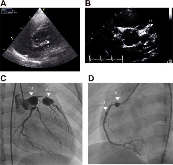
At the age of 3 years, he developed five major typical symptoms on the 7th day of the illness and was diagnosed with KD. He received intravenous immunoglobulin (IVIG) at local hospital and became afebrile on the 8th day of the illness. Although echocardiography was performed in the acute phase (Fig. 1A), after 1 and 3 months, CAAs were not diagnosed. Afterward, he did not return to the hospital at his parents’ decision.
At the age of 6 years, he underwent echocardiography as the 2nd examination of the SCS (Fig. 1B) because he was not regularly followed despite the apparent history of KD, and CAAs (diameters, 7.4 mm at segment 1, 9.1 mm at segment 5, and 9.5 mm at segment 6) were found. He was sent to the local hospital where giant CAAs at segment 1, 5, and 6 and 99% stenosis at segment 6 were demonstrated by contrast computed tomography, and oral aspirin and warfarin therapies were started. Because stress technetium myocardial perfusion scintigraphy using adenosine showed perfusion defect on the anterior septal wall, he was referred to our hospital for possible percutaneous coronary intervention.
Findings on Admission
His height was 120 cm, and weight was 19.6 kg. Heart rate was 93 bpm, and blood pressure was 112/60 mmHg. Cardiac examination on admission revealed regular rate and rhythm with normal S1 and S2 without S3 or murmurs. There were no abnormal findings in the abdomen and extremities and blood chemistry test.
Chest radiography showed cardiothoracic ratio of 40% without signs of calcification of the CAA. The 12-lead electrocardiogram showed normal sinus rhythm without ST-T segment change or abnormal Q wave. Echocardiography showed normal left ventricular dimension with normal ejection fraction of 72% without local asynergy, but CAAs of 9.1 mm and 9.5 mm in diameter were noted at segments 5 and 6 without thrombus.
Management and Outcome
Cardiac catheterization and coronary angiography (Fig. 1C and D) confirmed CAAs with maximum diameters of 10.8 mm, 12.3 mm, and 6.6 mm at segments 5, 6, and 1, respectively, and 99% stenosis between the CAAs of segments 5 and 6. After plain old balloon angioplasty was performed at the stenotic lesion and stenosis was successfully improved, follow-up coronary angiography after 8 months of angioplasty showed asymptomatic complete occlusion of giant CAA at segment 6. Because stress technetium myocardial perfusion scintigraphy using adenosine induced chest pain and obvious ST segment depression in V2–4, the patient was scheduled to undergo coronary artery bypass grafting and underwent a special treatment program with regular heparin infusion and exercise.4) Regular echocardiography showed suspected stenosis on the proximal side of the CAA at segment 1. The patient was transferred to another hospital where he underwent successful coronary artery bypass grafting (left internal mammary artery to the left anterior descending artery). Since then, he recovered without further intervention.
A 13-year-old boy in the 7th grade was selected for SCS because of a history of fever of unknown origin for >5 days.
Past Illness
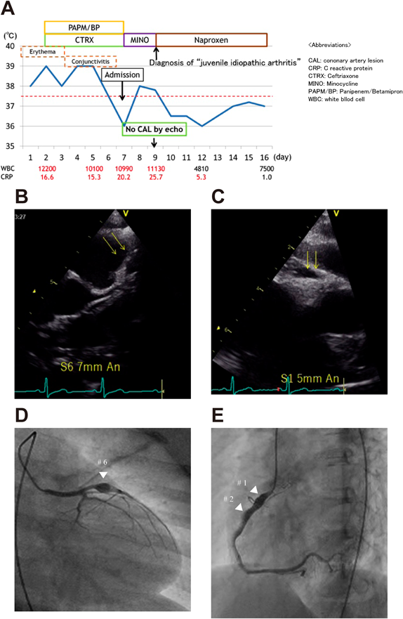
At the age of 9 years, he had fever and erythema of unknown origin that lasted for 9 days. Several antibiotics were ineffective, and he was diagnosed with juvenile idiopathic arthritis and received oral naproxen on day 9 of the illness that improved fever the next day (Fig. 2A). Throughout the course, the patient developed only fever, erythema, and joint pain. CAAs were not detected on echocardiography at day 9 of the illness, and thereafter echocardiography was not performed.
At the age of 13 years, the patient underwent echocardiography as the 2nd examination of SCS because he has a history of fever of unknown origin for >5 days, and CAAs (7 mm in diameter at segment 6 and 5 mm in diameter at segment 1) were found (Fig. 2B and 2C). He was admitted to our hospital for further examination. There was no unusual medical or family history.
Findings on Admission
His height was 144.2 cm, and weight was 28.9 kg. Heart rate was 74 bpm, and blood pressure was 108/80 mmHg.
Cardiac examination on admission revealed regular rate and rhythm with normal S1 and S2 without S3 or murmurs. There were no abnormal findings in the abdomen and extremities and in the blood chemistry test. Chest radiography showed a cardiothoracic ratio of 51% without signs of calcification of coronary aneurysm. The 12-lead electrocardiogram showed normal sinus rhythm without ST-T segment change or abnormal Q wave. Echocardiography revealed normal left ventricular dimension with normal ejection fraction of 64% without local asynergy and CAAs of 5 mm and 7.2 mm in diameter at segments 1 and 6, respectively, without thrombus.
Management and Outcome
The patient underwent cardiac catheterization and coronary angiography that confirmed CAAs at segment 6, proximal and distal parts of segment 1, and segment 2 (diameter 6.5 mm, 5.1 mm, 4.8 mm, and 3.9 mm, respectively) (Fig. 2D and 2E). There was no stenosis or thrombus. Since then, he has received oral aspirin therapy and recovered without any cardiac event.
We report two cases of CAAs that were not previously diagnosed during acute illness but were detected during SCS. These cases illustrate that SCS may provide an important opportunity to detect CAA in patients with a history of complete or incomplete KD. Guidelines for quality control and proper timing of echocardiography in patients with KD are required.
The reasons for the failure in detecting CAAs during acute illness include poor accuracy and inappropriate timing of echocardiographic examination and a lack of awareness that CAAs can develop even in patients with successful IVIG treatment.
In Case 1, the quality of echocardiographic examination during acute illness seems to be poor. The standard echocardiographic method5) is recommended for visualization and evaluation of the coronary artery in children. However, presently, there are no qualification criteria for those who evaluate the coronary artery during the acute phase of KD, such as physicians or ultrasonic technicians, and echocardiographic examinations have sometimes been performed using empirical and nonstandardized methods in hospitals where pediatric cardiologists do not work full time. Therefore, it is necessary to increase the number of physicians or technicians who have standardized skills to evaluate coronary arteries and maintain their skill levels through educational seminars possibly held by certain authorities, such as the Japan Society of Ultrasonics in Medicine.
Furthermore, Case 1 responded to a single dose of IVIG, and the examiner might have presumed that CAA would not be present. However, a certain number of single-dose IVIG responders have developed CAAs even though IVIG nonresponders are more likely to develop CAAs. While in echocardiographic examination, we must keep this fact in mind.
In Case 2, the patient had no history of persistent fever or hospitalization except for 9-day fever at the age of 9 years, but CAAs were noted for the first time at the SCS at the age of 13 years. In children, several diseases such as KD, chronic active Epstein–Barr virus infection,6) and meningococcal infection7) reportedly cause CAAs. In cases of juvenile idiopathic arthritis, coronary artery enlargement is observed in approximately 40% of patients, which is transient and does not progress to CAA.8) Therefore, considering the clinical courses, the patient seemed to have incomplete KD at the age of 9 years. The reason that the previous examiner did not detect CAA during acute illness is either poor accuracy or improper timing of echocardiographic examination. In Case 2, echocardiographic examinations were not performed after the 9th day of the illness, while he had been receiving treatment for juvenile idiopathic arthritis. Because CAAs in KD are often observed after day 10 of the illness,2) echocardiographic examinations for patients with suspected KD must be performed again within 1 month from the disease onset to rule out the development of CAAs.
We report two cases with CAAs that were not previously diagnosed during acute illness but were found for the first time during SCS. Quality control of echocardiography is required in patients with KD and suspected KD. In patients with suspected KD, it is necessary to perform echocardiography again at least 1 month after onset to check if CAAs are present.
Conflicts of Interest
The authors have no competing interests to declare.
Author Contributions
Takuro Kamura drafted the manuscript; Shintaro Kishimoto, Yoshiyuki Kagiyama, Hironaga Yoshimoto, and Yoshiyuki Kudo made the diagnosis, managed the patients, and co-drafted the manuscript; Kenji Suda revised the manuscript and made substantial scientific contributions.
All authors have read and approved the final version of the manuscript.
Note
This paper was partially presented at The 13th Study Group of Kawasaki Disease (May 31, 2014 in Kagoshima), and The 34th Annual Meeting of the Japanese Society of Kawasaki Disease (November 1, 2014 in Tokyo).
Originally published in Pediatric Cardiology and Cardiac Surgery, Vol. 33 (2017), No. 4, pp. 335–340
引用文献References
1) Research Committee of the Japanese Society of Pediatric Cardiology; Cardiac Surgery Committee for Development of Guidelines for Medical Treatment of Acute Kawasaki Disease: Guidelines for medical treatment of acute Kawasaki disease: Report of the Research Committee of the Japanese Society of Pediatric Cardiology and Cardiac Surgery (2012 revised version). Pediatr Int 2014; 5: 135–158
2) JCS Joint Working Group: Guidelines for diagnosis and management of cardiovascular sequel in Kawasaki disease (JCS 2013) digest version. Circ J 2014; 78: 2521–2562
3) Austen WG, Edwards JE, Frye RL, et al: A reporting system on patients evaluated for coronary artery disease. Report of the Ad Hoc Committee for Grading of Coronary Artery Disease, Council on Cardiovascular Surgery, American Heart Association. Circulation 1975; 51 Suppl: 5–40
4) Tateno S, Terai M, Niwa K, et al: Alleviation of myocardial ischemia after Kawasaki disease by heparin and exercise therapy. Circulation 2001; 103: 2591–2597
5) Fuse S, Kobayashi T, Arakaki Y, et al: Standard method for ultrasound imaging of coronary artery in children. Pediatr Int 2010; 52: 876–882
6) Nakagawa A, Ito M, Iwaki T, et al: Chronic active Epstein-Barr virus infection with giant coronary aneurysms. Am J Clin Pathol 1996; 105: 733–736
7) Ford SR, Rao A, Kochilas L: Giant coronary artery aneurysm formation following meningococcal septicaemia. Pediatr Cardiol 2007; 28: 300–302
8) Binstadt BA, Levine JC, Nigrovic PA, et al: Coronary artery dilation among patients presenting with systemic-onset juvenile idiopathic arthritis. Pediatrics 2005; 116: e89–e93
9) Ayusawa M, Sonobe T, Uemura S, et al: Kawasaki Disease Research Committee: Revision of diagnostic guidelines for Kawasaki disease (the 5th revised edition). Pediatr Int 2005; 47: 232–234