Journal of Pediatric Cardiology and Cardiac Surgery

Online ISSN: 2433-1783 Print ISSN: 2433-2720
Japanese Society of Pediatric Cardiology and Cardiac Surgery
Japanese Society of Pediatric Cardiology and Cardiac Surgery Academy Center, 358-5 Yamabuki-cho, Shinju-ku, Tokyo 162-0801, Japan
Journal of Pediatric Cardiology and Cardiac Surgery 3(2): 63-79 (2019)
doi:10.24509/jpccs.190201

ReviewReview

Recognition and Management of Preclinical Cardiomyopathies in Children: Special Focus on Duchenne Muscular Dystrophy and Anthracycline Cardiotoxicity

Nemours Cardiac Center, Nemours/Alfred I. duPont Hospital for Children ◇ Wilmington, DE 19803, USA

受付日:2019年3月20日Received: March 20, 2019
受理日:2019年5月17日Accepted: May 17, 2019
発行日:2019年7月1日Published: July 1, 2019
HTMLPDFEPUB3

Heart failure is conventionally defined as a dynamic clinical syndrome where the cardiovascular system fails to meet the metabolic demand of the peripheral tissues or can only do so at the expense of increased ventricular filling pressure. Emerging new findings in biomedical research have enabled us to recognize the early preclinical myocardial changes at the cellular and molecular levels before ventricular dysfunction becomes visible. These preclinical changes have also become recognizable by advanced medical technology. Here, we introduce two relatively common clinical entities that present with an asymptomatic preclinical stage in pediatrics, cardiomyopathies in Duchenne muscular dystrophy and anthracycline-induced cardiotoxicity. We will discuss underlying pathobiology, early diagnosis by noninvasive imaging studies, and cardioprotective treatment of the preclinical stage of progressive myocardial disease. The early recognition and treatment of the preclinical stage of heart failure may provide us with new perspectives in understanding this devastating disease.

Key words: preclinical heart failure; noninvasive imaging; cardiomyopathy; muscular dystrophy; cardio-oncology

Introduction

Heart failure (HF) has traditionally been defined as a hemodynamic abnormality in which the oxygen delivery through the circulation does not meet the metabolic demand of the peripheral organs or only does so at the expense of increased ventricular filling pressure. Recent advances in noninvasive imaging technology, most notably echocardiography and magnetic resonance imaging (MRI), have enabled us to identify an asymptomatic, early stage of HF with preserved ventricular systolic function. The recognition of this pre-HF stage is important, as initiation of early treatment may result in better outcomes than starting treatment when the disease is clinically established.1) Moreover, identifying the preclinical stage before the emergence of visible ventricular dysfunction may be of particular importance, as most HF patients present with an insidious stage followed by a progressive and potentially lethal clinical course.

In the following review, we focus on the underlying pathobiology of early evolution of HF, the clinical significance of preclinical HF, the diagnosis of preclinical HF with noninvasive imaging, and the treatment of preclinical cardiomopathies, especially those seen in Duchenne muscular dystrophy (DMD) and anthracycline-induced cardiotoxicity (AIC). Heart failure in congenital heart disease, valvular heart disease, and ischemic heart disease is not discussed.

Recognition of Preclinical Cardiomyopathies

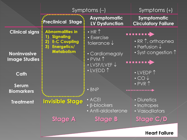
A classification of chronic HF in adults was published in 2005 by the American Heart Association (AHA)/American College of Cardiology (ACC)2) (Table 1). In this classification, Stage A was defined as a “preclinical” stage, where certain functional deterioration at the cellular level is present but is not enough to cause “visible” morphological changes, including ventricular dysfunction or dilatation. This stage inevitably progresses to Stage B, where tissue- or organ-level abnormality is commonly detected as ventricular dysfunction or dilatation by non-invasive imaging studies but without HF symptoms. Symptoms of HF arise once the low cardiac output persists and/or ventricular filling is compromised to induce venous congestion (Stage C) (Fig. 1). Multiple organs, including kidney, lung, liver, brain, bone, and skeletal muscles, are involved in worsening of clinically overt HF, suggesting that the advanced HF (Stage D) is a disease of multiple organ failure as a consequence of disrupted inter-organ biological crosstalk.3, 4)

Table 1 ACC/AHA 2005 Guideline Update for Diagnosis and Management of Chronic Heart Failure in Adults2)
Stage AAt high risk of HF
Structural heart disease§ (−). Symptoms (−)
Stage BStructural heart disease§ (+). Symptoms (−)
Stage CStructural heart disease§ (+). Symptoms (+)
Stage DRefractory CHF requiring specialized interventions
 Patients with hypertension, atherosclerosis, diabetes, obesity, or metabolic syndrome or using cardiotoxins or FH of CM. § Ventricular dilatation or hypertrophy.
Journal of Pediatric Cardiology and Cardiac Surgery 3(2): 63-79 (2019)

Fig. 1 Clinical Stages of Heart Failure

Clinical stages of heart failure (HF). Clinical syndrome of HF begins with a preclinical stage (or Stage A) that should be acknowledged as a “disease”. ACEI: antiogensin II-converting enzyme inhibitor, BNP: brain natriuretic peptide, CO: cardiac output, E-C coupling: excitation-contraction coupling, HR: heart rate, LV: left ventricular, LVEDD: LV end-diastolic dimension, LVEDP: LV end-diastolic pressure, LVEF: LV ejection fraction, LVSF: LV shortening fraction, PVM: pulmonary vascular markings, PVR: pulmonary vascular resistance, RR: respiratory rate, and Syst congestion: systemic congestion.

In the field of pediatrics, we encounter two relatively common patient populations with preclinical HF that are programmed to develop overt HF in a chronic phase. These include Duchenne muscular dystrophy (DMD) and anthracycline-induced cardiotoxicity (AIC). These two disorders recently have gained immense scientific interest because they serve as an excellent human model to understand the evolution of HF from preclinical to advanced stages with the availability of experimental animal models.5–8) The development of cardiomyopathy and clinical symptoms of HF is insidious but may progress to reach medically refractory HF (Stage D) in late teens in DMD9, 10) and early to mid-adulthood in AIC.11–13)

Duchenne muscular dystrophy is an X-linked genetic disorder that causes progressive weakness and wasting of skeletal muscle and myocardium in boys affecting one in 3,600 to 9,300 live male birth due to mutation of dystrophin.14, 15) The structural integrity of each individual skeletal and cardiac myocyte is significantly compromised upon physical stress due to absence of dytrophin,16–18) but the underlying mechanisms of muscle wasting are more complex, involving multiple different processes, including intracellular calcium overload,19–22) alteration in the nitric oxide (NO) pathway,23, 24) increased reactive oxygen species (ROS) and mitochondrial dysfunction,25–27) and extracellular matrix remodeling and myocardial fibrosis.28–31) There are certain other factors involved in the pathogenesis of DMD cardiomyopathy, including epigenetic factors, post-translational modification, telomere dysfunction, and genetic modifiers.32, 33) Dystrophin deficiency perturbs physiological myocyte growth in response to increased biomechanical stress as dystrophin serves as a mechanosensor that regulates protein synthesis via the NO pathway.24) Pathological alteration of DMD myocardium is heterogeneous34) and probably a combined consequence of myocardial wasting (myocyte loss or atrophy)35, 36) and secondary geometric changes (remodeling).37) The chamber dilatation and ventricular wall thinning occur in combination with secondary fatty infiltration and fibrosis upon myocyte death.38, 39) The earliest signs of cardiomyopathy in asymptomatic DMD are either decreased systolic function40) or resting sinus tachycardia.41) The mechanism of resting tachycardia is not well understood, but it is thought to be secondary to dysautonomia42) and/or decreased myocardial compliance due to fibrosis.41) The early clinical manifestation of HF is frequently overlooked, as most DMD patients usually become non-ambulatory and reliant on a wheelchair by age 13 because of progressive muscle weakness and wasting.37) Recent guidelines by American Heart Association suggested early prophylactic treatment with Angiotensin II-converting enzyme inhibitor (ACEi) before age 10 years regardless of left ventricular (LV) function.43)

Survivors of childhood cancer who received anthracycline therapy have a life-long risk of developing HF.11, 44–48) Several risk factors are known to affect the clinical course, including the cumulative dose of anthracycline, female sex, younger age at treatment, radiation therapy, genetic predisposition, and traditional risk factors for adult heart disease (hypertension, diabetes, smoking, hyperlipidemia, and obesity).49–51) The underlying mechanism of AIC is poorly understood but is thought to be multifactorial with complex mutual interactions,52–56) including increased ROS production,57, 58) intracellular calcium overload,59, 60) disrupted sarcolemmal integrity,61) mitochondrial dysfunction due to iron overload62) or enhanced autophagy,8) damaged DNA replication by topoisomerase 2β,63) impairment of cell survival pathways (neuregulin/ErbB signaling),64) premature senescence,65–67) and damage of cardiac stem cells.7, 66) These process are responsible for cardiomyocyte death and permanent myocardial injury through a common pathway of calcium overload, lipid peroxidation, and mitochondrial dysfunction,68) resulting in ventricular wall thinning, increased wall stress, ventricular dysfunction, and, eventually, DCM. Inadequate ventricular mass with chronic afterload excess may explain the persistent and progressive nature of AIC.12) One big mystery is the temporal gap between initial anthracycline exposure and initiation of cardiovascular adverse effects.52)

Based upon the onset, AIC is classified as a) acute (during or within 1 week of treatment: incidence <1%), b) early chronic (<1 year after treatment: ∼2%), and c) late-onset chronic (>1 year or a few decades after treatment: 5 to 57%).44, 51) Acute cardiotoxicity is relatively reversible, whereas late-onset chronic type is slowly progressive and irreversible that commonly leads to medically refractory HF. The incidence of cardiomyopathy in children who have received anthracycline-based chemotherapy is estimated at 3.3% at 10 years and 4.8% at 15 years,69) but the incidence of adverse cardiovascular events is higher with longer follow-up periods.48, 51, 70) The majority of the affected children remain asymptomatic with normal ventricular function during childhood, but the pathological process might have already been initiated at the molecular and cellular levels. As most of the late onset chronic cardiotoxicity presents during early to middle adulthood, it rarely catches the attention of pediatric medical providers unless there is an identifiable LV dysfunction. It is clear that detection of this preclinical phase gives us the best opportunity to impact the progression to the irreversible and life-threatening end-stage disease, at which point medical treatment has limited efficacy. The recommendations for primary surveillance to detect occult cardiotoxicity were published by Armenian and colleagues, optimizing the opportunities for collaborative research.71)

The underlying pathobiology of cardiomyopathy in DCM and AIC is summarized in Fig. 2. There are similarities between the two as myocyte death and subsequent wall thinning comprise a primary pathogenesis of both cardiomyopathies. In DMD cardiomyopathy, dysautonomia frequently causes persistent tachycardia, which increases myocardial oxygen demand. Because progressive myocyte loss and deficient LV mass predominates in DMD cardiomyopathy, ventricular dilatation may not be always evident even in the advanced stage. On the other hand, in AIC, cytotoxic effects target not only cardiomyocytes but also non-myocytes including endothelial cells, cardiac fibroblasts, and cardiac progenitor cells.11) Cardioprotective vascular development in response to ischemic injury and/or increased biomechanical stress may be mitigated, in part, due to disruption of paracrine crosstalk between the cells that plays a protective role against the adverse remodeling, responsible for an early onset of ischemic heart disease.64)

Journal of Pediatric Cardiology and Cardiac Surgery 3(2): 63-79 (2019)

Fig. 2 Pathobiology of cardiomyopathy (CM) in Duchenne muscular dystrophy (DMD) and anthracycline-induced cardiotoxicity (AIC)

Note that there are certain similarities between the two and that significant cellular and molecular abnormalities take place during Stage A heart failure (HF) in both conditions. In DMD, the progressive myocyte loss dominates over secondary ventricular remodeling, whereas the disruption of paracrine crosstalk among the cells mitigates intrinsic compensatory cardioprotective responses to biomechanical and ischemic stress in AIC. In DMD, ventricular remodeling (dilatation) does always take place because absence of dystrophin perturbs myocyte hypertrophy in response to increased shear stress. CFs: cardiac fibroblasts, CPCs: cardiac progenitor cells, DCM: dilated cardiomyopathy, ECs: endothelial cells, NO: nitric oxide, ROS: reactive oxygen species.

Diagnosis of Preclinical HF in Children

Echocardiography

Detecting subtle ventricular dysfunction and subclinical HF is a diagnostic challenge. Left ventricular systolic function can be assessed with measurement of shortening fraction (SF) on two-dimensional (2D) or M-mode short axis parasternal images. Shortening fraction simply reflects a change in LV chamber dimension during the cardiac cycle. SF %= ( LVEDDLVESD ) / LVEDD ×100

The Children’s Oncology Group adopted SF as a way of assessing the effect of chemotherapy on the heart. Shortening fraction is, however, limited in several ways: 1) it is obtained in one view and does not reflect motion of all of the cardiac segments; 2) it is unreliable in the setting of regional wall motion abnormalities, most notably changes in septal motion, which are common; and 3) it is affected both by preload and afterload.72, 73)

Ejection fraction (EF) has played a central role in the assessment of LV function in numerous adult clinical trials. Initial EF measurements were performed using gated radionuclide scans. Low EF measured with this technology was shown to correlate with increased mortality following myocardial infarction.74) Ejection fraction describes the change in volume during the cardiac cycle and is calculated as follows: EF % =(LV end diastolic volume [LVEDV] LV end systolic volume [LVESV])/LVEDV ×100

The numerator measures stroke volume (SV), which varies directly with EF. The denominator assesses the degree of dilation, which varies inversely with EF. Several formulas have been developed, taking into account the ellipsoid shape of the LV and using the linear chamber diameter, as measured for SF, to then calculate an estimate of EF.72, 73) One of the more commonly utilized approaches is the Teichholz method, which uses this equation: Volume [V]=7/ ( 2.4+diameter [D] ) × D 3 Volume is measured with this formula at end-diastole and end-systole, allowing calculation of SV and EF.

Currently, the American Society of Echocardiography (ASE) and the European Association of Cardiovascular Imaging (EACVI) recommend using the biplane Simpson’s method of multiple discs to calculate LV volumes and EF in adults.75) To accomplish this calculation, the LV endocardial border is traced in two orthogonal planes (typically apical four-chamber and apical two-chamber views or the equivalent transesophageal echocardiographic [TEE] views) at end-diastole and end-systole. A computer algorithm then divides the traced area into 20 discs of equal height and sums the volume of each disc. The endocardial tracing is performed automatically on most current generation echocardiography machines. Manual adjustment of the borders remains important to consistently exclude trabeculations, exclude noise in the near field (apex), and assess for fairly uniform wall thickness. The biplane Simpson’s EF does correlate with outcomes and mortality in adults.76) Limitations include poor endocardial definition due to limited acoustic windows, diminished reliability in the setting of arrhythmia and difficulty defining end-systole and end-diastole when there is significant dyssynchrony.73)

In pediatric patients, SF and EF have been shown to be associated with prognosis in DCM. In a study of 1,426 children with DCM undertaken between 1996 and 2003, independent risk factors for subsequent death or transplantation included older age, congestive HF, cause of DCM, and lower LVSF z score (p<0.001 for all).77) A meta-analysis of pediatric DCM by Alvarez et al. indicated that higher LV EF and SF at diagnosis were predictive of better prognosis across multiple articles. Younger age at diagnosis and the presence of myocarditis as the etiology were also associated with better prognosis.78) The outcomes of 1953 children with DCM over two decades by the Pediatric Cardiomyopathy Registry demonstrated that worse LV SF at diagnosis independently predicted death.79) Echocardiographic parameters of LVSF z score and LVEDD z score predicted a higher risk of sudden death in 289 children with DCM, HCM, LV noncompaction cardiomyopathy, and arrhythmogenic cardiomyopathy.80) While these measures are very useful in overt HF, they are not sufficiently sensitive to detect preclinical myocardial dysfunction. This is particularly true in pediatric patients given the challenges with reproducibility.

Assessment of diastolic function is important but challenging, particularly in children.81) Diastolic dysfunction typically accompanies overt systolic dysfunction but can occur independently in the setting of hypertrophic cardiomyopathy (HCM), restrictive cardiomyopathy (RCM), or in patients with HF preserved EF (HFpEF).82) Changes in diastolic function can also precede systolic dysfunction. Echocardiography is an important means of assessing diastolic dysfunction and utilizes the following parameters:

  1. 1) Transmitral Doppler inflow pattern: Early filling (E wave) and late filling (A wave) velocities are measured in an apical four chamber view, and E/A ratio is calculated. Moving to an apical five-chamber view and placing the sample volume midway between the mitral leaflet tips and the LV outflow tract, isovolumic relaxation time (IVRT), acceleration time (AT), and deceleration time (DT) are measured from the Doppler tracing.
  2. 2) Pulmonary venous Doppler flow pattern: The flow pattern at the right upper pulmonary vein is characterized by an initial S wave during systole, a D wave during diastole and an A wave, representing reversal of flow from the left atrium into the pulmonary veins during atrial systole.
  3. 3) Tissue Doppler imaging (TDI): TDI assesses myocardial motion rather than blood flow. The sample volume is placed within the myocardial tissue immediately adjacent to the septal and lateral hinge-points of the mitral valve. Early (E′) and late (A′) velocities can be measured. A ratio of the mitral inflow E velocity to the tissue Doppler E′ velocity is typically calculated (E/E′ ratio).
  4. 4) Color M-mode flow propagation velocity (Vp): This measurement is obtained by placing the M-mode cursor through the center of the mitral inflow jet in an apical four-chamber view. The velocity of propagation is the slope of the first aliasing velocity during early filling or the slope of the line where there is a transition from no color to color.

In children, the adult guidelines for assessment of diastolic function do not perform as well. In a 2013 study, three highly-respected investigators, Dragulescu, Mertens, and Friedberg, each measured parameters of diastolic function in 175 children, including patients with DCM, HCM, RCM and a control group. Despite elevated filling pressures in half of the patients, echocardiographic parameters were abnormal in only 14% of the HCM group and in none of the DCM and RCM groups. There was poor interobserver agreement on grading of the diastolic dysfunction using the adult algorithm. Using pediatric reference data rather than the adult data yielded slightly better results, classifying all of the controls as normal, but diastolic dysfunction was detected in only a small proportion of the patients despite severe disease.83)

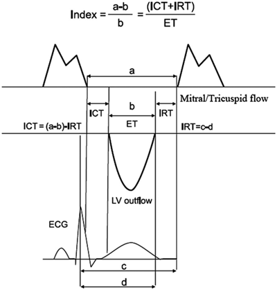
The myocardial performance index (MPI), or Tei index, assesses global ventricular function.84) It is obtained by dividing the sum of the isovolumic contraction time (ICT) plus isovolumic relaxation time (IVRT) by the ejection time (ET). In practice, it is easier to measure the interval from cessation to onset of mitral inflow (a) minus the ET (b) as a substitute for ICT+IRT (Fig. 3). The MPI can be measured on the mitral or tricuspid inflow Doppler or can be measured on tissue Doppler tracings. Compared with Doppler-derived MPI, TDI-derived measurements have the advantage of being less affected by loading conditions and heart rate and more sensitive to diastolic function. In one small study of 25 pediatric patients with HF, TDI-derived septal MPI was inversely correlated with SF and reflected severity of disease. As with SF and EF, MPI is most useful in patients with overt HF and less useful in detection of early or subclinical ventricular myocardial changes. A prospective study in 2010 compared the E/E′ ratio and MPI to near-simultaneous catheter derived hemodynamic parameters in post-transplant children. The correlations were poor.85)

Journal of Pediatric Cardiology and Cardiac Surgery 3(2): 63-79 (2019)

Fig. 3 Schematic presentation of myocardial performance index (Tei index)

ET: ejection time, ICT: isovolumic contraction time, and IRT: isovolumic relaxation time. For detail, please see the text.

A breakthrough in the ability to assess subtle ventricular dysfunction came with the development of strain echocardiography.86, 87) Strain is based on speckle-tracking technology, which follows a point on the myocardium as it moves through the cardiac cycle. This technology highlights the complexity of myocardial deformation; the ventricle shortens, thickens and twists during systole to eject blood. This angle-independent technique allows quantification of segmental and global ventricular function. Strain can be measured in longitudinal, circumferential and radial planes. The vast majority of data thus far are focused on global longitudinal strain (GLS), which is measured in apical four-chamber, three-chamber, and two-chamber views. Normal values for GLS range from −19% to −20%. In the adult literature, GLS is a valuable prognostic tool and an independent predictor of all-cause mortality in HF patients.88) In multiple studies of adult patients who have undergone chemotherapy, changes in GLS consistently precede significant changes in EF89) and predict cardiotoxicity.90) In a longitudinal study of 737 adult patients, lower (less negative) baseline GLS values predicted mortality in asymptomatic patients with primary mitral regurgitation and preserved EF.91)

Cardiac MRI

Cardiac MRI (CMRI) is currently the reference standard imaging modality for the quantification of ventricular volumes, mass, and EF.92, 93) It has high temporal and spatial resolution in the assessment of RV and LV volume, systolic function, and mass with high interobserver and intraobserver reproducibility.94–96) In contrast to echocardiography, CMRI can obtain high-resolution cine images of the heart in any imaging plane without the limitations of acoustic windows. Normal values are now available on RV and LV volume, mass, and EF for different age groups in males and females, including children.97) Steady-state free precession (SSFP) cine imaging is the current standard method for measuring volumetry in CMRI, replacing cine gradient echo sequences. Cardiac MRI is increasingly used for evaluation, management, and prognosis of patients with HF.92, 93) The method of discs is used for quantification of RV and LV end diastolic and end systolic volumes, and EF can be calculated without making geometric assumptions; this makes CMRI suitable even with dilated and remodeled ventricles.98) In adult patients with HF, EF is a well-recognized factor in the long-term prognosis and survival.99) In the pediatric population, limited data have shown that indices of LV systolic performance such as EF and SF are major determinants of prognosis, in addition to age, presence of congestive HF, and LV dimension z score.100)

Late gadolinium enhancement (LGE) is an MRI sequence that uses gadolinium (Gd) contrast to evaluate for fibrosis in the ventricular myocardium. Gadolinium distributes into the extracellular space of the myocardium but not into the normal myocardial cells.101, 102) In areas of myocardial cell necrosis, there is an increase in Gd distribution into the cells with loss of cell membrane integrity and into the expanded extracellular matrix.103) The increased Gd results in shortened T1 values of the affected areas that results in increased signal intensity on special T1-weighted MRI sequences, such as inversion recovery gradient echo images.104) In adult patients with nonischemic DCM, Assomull et al. showed that midwall fibrosis was present in about 35% of patients and was a predictor of death or hospitalization for cardiovascular events (hazard ratio 3.4) in addition to sudden cardiac death and ventricular tachycardia (hazard ratio 5.2), independent of other factors such as EF.105)

More recent and newer CMRI techniques have emerged to quantify fibrosis in cardiomyopathies in addition to providing a more accurate volume assessment than echocardiogram. These techniques examine the tissue characteristics of the myocardium and quantify the extracellular volume, which is increased in myocardial fibrosis. One such method is T1 mapping of the myocardium both before and after the use of Gd contrast. Animal models106) and patients post-heart transplant and with DCM107) showed correlation between T1 mapping and myocardial fibrosis. Another technique used in CMRI is strain measurement. One such sequence uses MRI tagging, where a grid is created by MR pulses and the deformation measured in systole, but the principal image tracking method is different between CMRI and echocardiography.108)

Diagnostic Applications by Non-Invasive Imaging Studies

Duchenne Muscular Dystrophy

The primary pathological process in DMD cardiomyopathy is progressive myocyte loss with perturbed compensatory growth response and resultant myocardial fibrosis (Fig. 2). Significant changes in SF and EF tend to occur during adolescence in DMD patients. With the advancement in respiratory support of this group of patients and their longer survival, cardiomyopathy has become the leading cause of death.109) Due to limited mobility of DMD patients, overt symptoms of congestive HF are not recognized until the advanced stages of cardiomyopathy.

Ryan et al. measured SF and circumferential strain by transthoracic echocardiogram in a group of young (<8 years of age) patients with DMD and a control group and reported significant changes in the circumferential strain values in the DMD group compared with the control group at mid-ventricular anteroseptal, inferior, and infero-lateral segments, which could be a marker of subclinical cardiac disease in this group of patients starting in the first decade of life.110) Markham et al. compared diastolic function parameters between patients with DMD (<15 years of age with no HF) and controls. They assessed different diastolic function parameters, including Doppler-derived mitral E/A ratio, tissue Doppler-derived E′/A′ ratio, E/E′ ratio, IVRT, and mitral valve deceleration time. In this study, DMD had significantly worse findings overall on their diastolic function indices; patients who developed HF at follow-up had even worse diastolic function parameters at initial assessment compared with those who continued to have no manifestations of HF, signifying the importance of diastolic function abnormalities as a marker for future development of HF in young DMD patients.111) Another study by Spurney et al. showed that patients with DMD had significantly higher mitral A-wave velocities, lower E/A ratios, and lower Doppler tissue imaging mitral lateral E′ velocities when compared with the age-matched control subjects, indicating impaired diastolic function. In addition, the same study revealed that speckle-tracking derived myocardial strain and strain rate values (both longitudinal and circumferential) were significantly different in the DMD group compared with controls. The differences were notably significant, even in patients younger than 13 years with normal SF and normal MPI values.112)

Strain analysis in 19 DMD patients and 16 controls revealed significantly lower global circumferential strain (GCS) (−14.7±4.7 vs −23.1±2.9, p=0.001) and GLS (−13.6±5 vs −18.8±3, p=0.001) than the controls, suggesting that occult cardiovascular dysfunction was present.113) Circumferential strain was reduced in DMD patients compared with controls, even at a young age when other systolic function parameters were still normal, and worse strain values were noted in patients with decreased EF.114) Similar results were obtained by Ashford et al. in DMD patients.115) Hagenbuch et al. studied circumferential strain in DMD patients over a mean period of 15 months and found progressive decrease in strain values over time in all patients.116) Strain and strain rate have also detected early changes in cardiac function in adolescent patients with type 1 diabetes, in asymptomatic children with LV noncompaction and normal EF, and in heart transplant patients during an episode of rejection.117–119) While strain is clearly promising for the detection of subclinical and mild myocardial dysfunction, challenges include reproducibility of measurements and intervendor differences.120)

The diagnostic value of CMRI has been emphasized in detection of preclinical HF in DMD. In a small study involving 10 patients (ages 7 to 18 years) with DMD or Becker muscular dystrophy (BMD), Silva et al. investigated myocardial fibrosis by LGE-CMRI. Fibrosis was found in 70% of the patients tested, and those patients with fibrosis had significantly lower LVEF and higher LVESV.39) Of note, only two patients had abnormal findings on echocardiogram, signifying the importance of LGE in early detection of cardiomyopathy in muscular dystrophy. Soslow et al. compared T1 values after Gd contrast administration between patients with DMD and controls and found that DMD patients had shorter post-contrast T1 values compared with controls, even those patients without LGE or reduced EF.121) In a larger study on patients with DMD, Tanden et al. retrospectively reviewed the association of LGE to LVEF, rate of decline of EF, and effect of treatment with steroids.122) This study included 98 patients with DMD who had four or more CMRI studies finding that the age of onset of LV dysfunction was variable. This study showed that the rate of decline of LVEF in these patients increased after development of fibrosis on LGE imaging, and EF decline correlated with number of segments affected. They also found that longer duration of steroid treatment may decrease the rate of new onset fibrosis.122) Studies in adults with ischemic heart disease showed that LGE on CMRI is a better predictor of inducible monomorphic ventricular tachycardia than EF123) and development of major cardiac events and cardiac mortality.124) More studies are needed to evaluate the impact of LGE in DMD on prognosis and outcome. The limitations of LGE include the semiquantitative nature of the sequence and limited ability to assess for scarring in the presence of diffuse myocardial fibrosis.

Anthracycline-Induced Cardiotoxicity

Among those who survive their malignancy, cardiotoxicity is the third leading cause of morbidity and mortality, following relapse of primary malignancy and occurrence of secondary cancer.125) Central pathogenesis of AIC may be outlined by direct cardiomyocyte impairment causing myocardial thinning and ventricular dysfunction and premature senescence affecting endothelial cells and other cell types (Fig. 2). Cardinale et al. showed, in an adult study of AIC, that earlier treatment of HF is associated with a more favorable response to HF therapy and lower rate of cardiac complications.126) Earlier studies showed that pediatric patients who received anthracycline chemotherapy demonstrated acute decrease of systolic and diastolic function measurements immediately after chemotherapy; although the values remained within normal limits.127) However, the decline in LV systolic function is not a sensitive or reliable marker for early myocardial deterioration by AIC.128) It is essential to detect AIC in the preclinical stage to prevent long-term irreversible adverse cardiomyopathy.

Mavinkurve-Groothuis et al. showed that all myocardial strain parameters decreased during chemotherapy and the degree of decrease was proportional to the cumulative anthracycline dose despite normal SF in these patients. The changes in the strain parameters persisted when other biomarkers of cardiac damage such as troponin T level did not predict abnormalities in the strain.129) In one study, 71 children exposed to anthracycline chemotherapy were found to have diminished longitudinal and circumferential echocardiographic strain and particularly diminished diastolic strain rate despite having normal SF.130) Cetin and colleagues measured conventional echocardiographic parameters and strain in 45 childhood anthracycline-treated cancer survivors and age-matched controls. Both groups had normal EF, SF, and brain natriuretic peptide levels; the anthracycline group had significantly lower longitudinal and radial strain values.131) Poterucha et al. compared two-dimensional speckle-tracking echocardiography-derived longitudinal peak systolic strain (LPSS) values in adolescent patients receiving anthracycline chemotherapy and demonstrated small but significant changes in LPSS values at 4 and 8 months post-chemotherapy, compared with pre-chemotherapy and controls, that preceded the decline in EF.132) Comprehensive echocardiographic assessment, including three-dimensional (3D) LVEF, global longitudinal and circumferential myocardial strain, and diastolic function in 1820 adult survivors of childhood cancer, revealed that abnormal GLS and diastolic function were more prevalent than reduced 3D LVEF.128) However, current guidelines for AIC surveillance are heavily dependent upon 2D echocardiographic assessment of LV systolic performance as a primary method.71, 133) The advanced echocardiographic methods may be incorporated into these guidelines.

Toro-Salazar et al. demonstrated that CMRI-derived abnormal myocardial characteristics and abnormal regional myocardial function were detected in pediatric cancer survivors with normal LV systolic function, suggesting that CMRI parameters, LV peak circumferential strain, T1 mapping-derived relaxation time, and extracellular volume (ECV), were effective in detecting occult AIC.134) In comparison with echocardiogram, CMRI not only demonstrated higher incidence of reduced EF among 114 adult survivors of childhood cancer, but also more frequently identified preclinical LV dysfunction in previously undiagnosed survivors.135) Tham et al. studied late cardiotoxic effects in 30 pediatric cancer survivors (age 15±3 years) and demonstrated that myocardial T1 values and ECV correlated well with cumulative anthracycline dosage, exercise capacity, and LV remodeling.136) On the other hand, in 91 adult cancer survivors with reduced EF (35±8%), CMRI-derived indexed LV mass showed strong correlation with the total anthracycline dosage and the incidence of major adverse cardiovascular events, but the incidence of myocardial fibrosis by LGE was infrequent in this study.137) Further studies are warranted to reliably identify the preclinical stage of AIC by noninvasive imaging modalities.

Management of Preclinical HF

Preclinical HF in the pediatric population has not been well studied and unfortunately is not recognized until it progresses to an advanced stage with clinically overt symptoms. Guidelines from both European and United States cardiology societies do not provide specific recommendations for management of preclinical HF.138, 139) The ultimate goal of HF therapy is to improve hemodynamics, prevent disease progression, maintain quality of life, and allow optimal growth and development in these children, but currently available conventional HF therapy has very limited capacity. Most of the current recommendations are extrapolated from adult guidelines, as we commonly see in pediatrics. Within the field of pediatric HF, angiotensin-converting enzyme inhibitors (ACEi) are considered first-line of therapy and β-blockers are second-line. Anti-mineralocorticoids are also accepted in the treatment of pediatric HF,140) whereas diuretics are recommended only to be used to achieve a euvolemic status.141)

Duchenne Muscular Dystrophy

Cardiac dysfunction is a well-known and important complication of some neuromuscular disorders, especially in DMD.37, 43, 142–145) The cardiac status of DMD patients is difficult to assess clinically given their musculoskeletal and respiratory limitations. A study published in 2005 evaluated the effect of perindopril in patients with DMD with normal baseline EF, which demonstrated that early treatment, before the presence of symptoms and between the ages of 9 and 13 years, can delay the onset and progression of clinically-apparent LV dysfunction in children with DMD.146) Thus, recognition of the preclinical stage and early treatment appear critical for favorable outcome.

Cardioprotective treatment of preclinical cardiomyopathy in DMD has been widely advocated in recent years.10, 147) Previous studies have shown some benefit from ACEi in patients with DMD.148, 149) A study led by Viollet showed overall improvement in LV function in patients treated, but there was no significant difference between patients treated with an ACEi alone or in combination with a β-blocker.150) A small, randomized, double-blind trial of lisinopril or losartan in 22 patients showed equal effectiveness in improving LVEF over a year of observation by echocardiogram.151) β-blockers are reported to be beneficial when used in combination with ACEi.152) Despite the lack of strong evidence, the International Society of Heart and Lung Transplantation (ISHLT) expert panel recommended on their guidelines that ACEi therapy should be considered for DMD unless there is a specific contraindication153); although the optimal age of institution of therapy is not clearly delineated.

Since the benefit of preventive therapy has not been clearly proven, it is challenging to decide the timing to initiate cardiac pharmacotherapy in this complex and heterogeneous population.154) On the other hand, oral corticosteroids are commonly prescribed early with the goal of preserving skeletal muscle strength and pulmonary function.155) Multiple mechanisms have been proposed for the beneficial biological role of steroids in DMD myopathy, including nuclear effect via activation of calcineurin/nuclear factor of activated T cells (NF-AT) pathway, enhancement of proliferation of myogenic precursor stem cells, reduction in the rate of muscle breakdown, and direct transcriptional modifiers to increase synergistic molecules, i.e., utrophin.156) A study evaluating 462 boys with DMD concluded that patients treated with corticosteroids can have a delayed onset of cardiac dysfunction and up to a 20% lower likelihood of developing cardiomyopathy compared with untreated patients.157) Schram et al. demonstrated a positive impact of steroids in mitigating the development of DCM and mortality in 87 patients (mean age 9.1±3.5 years) followed for more than a decade.158) However, significant adverse effects of steroids need to be closely monitored.

A comprehensive and multidisciplinary approach is required to provide patients with optimal prevention, monitoring, and treatment strategies. Currently, a randomized, multi-center, placebo-controlled clinical trial has been initiated in the United Kingdom to investigate whether the introduction of ACEi (perindopril) and β-blocker (bisoprolol) before the onset of identifiable LV systolic dysfunction can attenuate the development of cardiomyopathy in DMD patients aged 5 to 14 years.159)

Anthracycline-Induced Cardiotoxicity

Chemotherapy-induced cardiotoxicity, most commonly caused by anthracyclines, can be silent initially but take years or decades to manifest as cardiomyopathy, HF, myocardial infarction, myocarditis, or other significant clinical cardiac dysfunction.70, 160) There is no consensus regarding prevention or treatment of asymptomatic patients with AIC, and patients with cancer are often excluded from large HF trials.161) The underlying mechanisms of AIC are poorly understood, but there are a number of pathological processes proposed by multiple investigators, as discussed earlier.

Various agents have been proposed for the prevention of cardiomyopathy due to AIC.56) A meta-analysis by Kalam et al. showed that different agents such as dexrazoxane, statins, ACEi, and β-blockers may all have a cardioprotective effect.162) Dexrazoxane prevents AIC not only by chelating redox-active iron and subsequently reducing ROS formation, but also by reducing anthracycline-induced DNA damage via topoisomerase 2-α and topoisomerase 2-β.163) Cardinale et al. reported the benefits of ACEi in patients treated with anthracyclines.126, 164) In adult patients with acute myocardial cell injury after high-dose chemotherapy, early treatment with ACEi demonstrated significant protective effects against LV systolic dysfunction (LVEF: 62.4±3.5% and 48.3±9.3% in ACEi and non-treated group, respectively).164) Complete recovery of the LV systolic function was observed in 64% of the patients when ACEi was initiated soon after therapy, whereas the cardioprotective effect was significantly diminished after 12 months of the chemotherapy.126) A well-designed double-blind, placebo-controlled study was conducted with the aim to test if ACEi would decrease the LV end-systolic wall stress (LVESWS) and therefore improve or prevent cardiac dysfunction.165) The study showed decreased LVESWS by ACEi, but the reduction was not associated with significant changes on LVEF or LVSF, and there were more side effects in the treated than the untreated group. The most recent pediatric HF guidelines from ISHLT have no specific recommendation for ACEi therapy in stage A HF, whereas they recommend routine use of ACEi for the treatment of asymptomatic LV dysfunction (HF Stage B) unless there is a specific contraindication.153) The decision to extrapolate these results and routinely treat patients with subclinical HF with ACEi requires careful consideration given possible side effects that could outweigh potential benefits.

Initial studies have shown that β-blockers can provide a significant cardioprotective effect on both LV systolic and diastolic function.166, 167) The cardioprotective effects of β-blockers are thought to be due to increased pro-survival signaling via recruitment of β-arrestin and transactivation of epidermal growth factor receptor (EGFR), mitigation of oxidative stress, and prevention of myocardial calcium overload, resulting in enhanced lusitropy,56) in addition to reduction of myocardial workload through negative chronotropic and inotropic effects. Carvedilol, a non-selective β-blocker, has been used to treat cardiotoxicity and has improved LV function and mortality in adults. The evidence in pediatrics is much more limited. A small prospective randomized control trial, called the OVERCOME trial showed an absolute difference of 3.1% in LVEF in patients being treated with carvedilol and enelapril compared with placebo.168) Other studies have shown a similar protective effects in patients treated with β-blockers.161, 169) One could recommend a slow titration of this therapy, but the evidence remains limited.

There is still no clear evidence to define whether prophylactic cardioprotection is effective in preventing progression of cardiomyopathy in pediatric cancer survivors, or whether healthy life style and exercise can help prevent myopathic changes in the heart. Although there are no published large human clinical trials, evidence from numerous animal studies suggests that aerobic exercise training is protective against AIC.170, 171) In a small prospective study in 21 long term childhood acute lymphoblastic leukemia survivors with normal EF, home-based exercise improved diastolic function indicated by TDI and peak circumferential systolic and diastolic strain rates.172) Aerobic exercise may modulate cardiotoxicity by mitigating oxidative stress, pro-apoptotic signaling, ultrastructural changes secondary to calcium overload, and energy metabolism alteration, and by enhancing protein synthesis.173, 174) While counseling healthy lifestyle for these patients is warranted, there is a need for new trials that assess the effects of healthy diet, exercise, and prophylactic cardiac medical therapies on cardiac morbidity and mortality in this high-risk population.

Future Perspectives

The study of preclinical HF in the pediatric population has been limited, primarily because the patients are asymptomatic and because traditional assessment of ventricular function misses the early changes. Cardio-oncology is a new but under-recognized field in the pediatric cardiology that warrants further endeavor in understanding of epidemiology, screening, prevention, and treatment of the disease.175) Furthermore, conventional HF therapies may improve symptoms but not cure the underlying pathologic processes. This paradigm needs to change in light of recent advances in technology that make this previously “invisible” phenomenon “visible”. At the same time, evolution of scientific research has helped us understand the cellular and molecular level of myocyte pathobiology and the numerous communications between cardiomyocytes, fibroblasts, endothelial cells, circulating monocytes, and cardiac stem cells. A broad concept of HF should be classified into further specific subgroups based upon the underlying pathobiology, and the treatment of HF should follow accordingly. The concept of “precision medicine” needs to be introduced in pediatric HF.176) Duchenne muscular dystrophy and AIC provide excellent human models to study the evolution of HF from the preclinical to the advanced stage. Enhanced collaborative efforts between clinical medicine and basic science research are essential to understand the very mechanism of pathological transformation of myocardial tissue and to determine strategies to effectively halt or slow the progression.

Funding

None applicable.

Conflicts of Interest

None.

引用文献References

1) Wan SH, Vogel MW, Chen HH: Pre-clinical diastolic dysfunction. J Am Coll Cardiol 2014; 63: 407–416

2) Hunt SA, Abraham WT, Chin MH, et al: American College of Cardiology, American Heart Association Task Force on Practice Guidelines, American College of Chest Physicians, International Society for Heart and Lung Transplantation, Heart Rhythm Society: ACC/AHA 2005 Guideline Update for the Diagnosis and Management of Chronic Heart Failure in the Adult: A report of the American College of Cardiology/American Heart Association Task Force on Practice Guidelines (Writing Committee to Update the 2001 Guidelines for the Evaluation and Management of Heart Failure): Developed in collaboration with the American College of Chest Physicians and the International Society for Heart and Lung Transplantation: endorsed by the Heart Rhythm Society. Circulation 2005; 112: e154–e235

3) Husain-Syed F, McCullough PA, Birk HW, et al: Cardio-pulmonary-renal interactions: A multidisciplinary approach. J Am Coll Cardiol 2015; 65: 2433–2448

4) Jahng JW, Song E, Sweeney G: Crosstalk between the heart and peripheral organs in heart failure. Exp Mol Med 2016; 48: e217

5) Deconinck AE, Rafael JA, Skinner JA, et al: Utrophin-dystrophin-deficient mice as a model for Duchenne muscular dystrophy. Cell 1997; 90: 717–727

6) Megeney LA, Kablar B, Perry RL, et al: Severe cardiomyopathy in mice lacking dystrophin and MyoD. Proc Natl Acad Sci USA 1999; 96: 220–225

7) Huang C, Zhang X, Ramil JM, et al: Juvenile exposure to anthracyclines impairs cardiac progenitor cell function and vascularization resulting in greater susceptibility to stress-induced myocardial injury in adult mice. Circulation 2010; 121: 675–683

8) Koleini N, Kardami E: Autophagy and mitophagy in the context of doxorubicin-induced cardiotoxicity. Oncotarget 2017; 8: 46663–46680

9) Hor KN, Mah ML, Johnston P, et al: Advances in the diagnosis and management of cardiomyopathy in Duchenne muscular dystrophy. Neuromuscul Disord 2018; 28: 711–716

10) D’Amario D, Amodeo A, Adorisio R, et al: A current approach to heart failure in Duchenne muscular dystrophy. Heart 2017; 103: 1770–1779

11) Chen MH, Colan SD, Diller L: Cardiovascular disease: Cause of morbidity and mortality in adult survivors of childhood cancers. Circ Res 2011; 108: 619–628

12) Lipshultz SE, Lipsitz SR, Sallan SE, et al: Chronic progressive cardiac dysfunction years after doxorubicin therapy for childhood acute lymphoblastic leukemia. J Clin Oncol 2005; 23: 2629–2636

13) Lipshultz SE, Colan SD, Gelber RD, et al: Late cardiac effects of doxorubicin therapy for acute lymphoblastic leukemia in childhood. N Engl J Med 1991; 324: 808–815

14) Emery AE: Population frequencies of inherited neuromuscular diseases: A world survey. Neuromuscul Disord 1991; 1: 19–29

15) Mah JK, Korngut L, Dykeman J, et al: A systematic review and meta-analysis on the epidemiology of Duchenne and Becker muscular dystrophy. Neuromuscul Disord 2014; 24: 482–491

16) Wallace GQ, McNally EM: Mechanisms of muscle degeneration, regeneration, and repair in the muscular dystrophies. Annu Rev Physiol 2009; 71: 37–57

17) Yasuda S, Townsend D, Michele DE, et al: Dystrophic heart failure blocked by membrane sealant poloxamer. Nature 2005; 436: 1025–1029

18) Rybakova IN, Patel JR, Ervasti JM: The dystrophin complex forms a mechanically strong link between the sarcolemma and costameric actin. J Cell Biol 2000; 150: 1209–1214

19) Fong PY, Turner PR, Denetclaw WF, et al: Increased activity of calcium leak channels in myotubes of Duchenne human and mdx mouse origin. Science 1990; 250: 673–676

20) Fanchaouy M, Polakova E, Jung C, et al: Pathways of abnormal stress-induced Ca2+ influx into dystrophic mdx cardiomyocytes. Cell Calcium 2009; 46: 114–121

21) Franco A Jr., Lansman JB: Calcium entry through stretch-inactivated ion channels in mdx myotubes. Nature 1990; 344: 670–673

22) Viola HM, Adams AM, Davies SM, et al: Impaired functional communication between the L-type calcium channel and mitochondria contributes to metabolic inhibition in the mdx heart. Proc Natl Acad Sci USA 2014; 111: E2905–E2914

23) Wehling-Henricks M, Jordan MC, Roos KP, et al: Cardiomyopathy in dystrophin-deficient hearts is prevented by expression of a neuronal nitric oxide synthase transgene in the myocardium. Hum Mol Genet 2005; 14: 1921–1933

24) Garbincius JF, Michele DE: Dystrophin-glycoprotein complex regulates muscle nitric oxide production through mechanoregulation of AMPK signaling. Proc Natl Acad Sci USA 2015; 112: 13663–13668

25) Zorov DB, Juhaszova M, Sollott SJ: Mitochondrial reactive oxygen species (ROS) and ROS-induced ROS release. Physiol Rev 2014; 94: 909–950

26) Williams IA, Allen DG: The role of reactive oxygen species in the hearts of dystrophin-deficient mdx mice. Am J Physiol Heart Circ Physiol 2007; 293: H1969–H1977

27) Khairallah M, Khairallah R, Young ME, et al: Metabolic and signaling alterations in dystrophin-deficient hearts precede overt cardiomyopathy. J Mol Cell Cardiol 2007; 43: 119–129

28) Dahiya S, Givvimani S, Bhatnagar S, et al: Osteopontin-stimulated expression of matrix metalloproteinase-9 causes cardiomyopathy in the mdx model of Duchenne muscular dystrophy. J Immunol 2011; 187: 2723–2731

29) Delfín DA, Zang KE, Schill KE, et al: Cardiomyopathy in the dystrophin/utrophin-deficient mouse model of severe muscular dystrophy is characterized by dysregulation of matrix metalloproteinases. Neuromuscul Disord 2012; 22: 1006–1014

30) Wynn TA: Cellular and molecular mechanisms of fibrosis. J Pathol 2008; 214: 199–210

31) Zhou L, Lu H: Targeting fibrosis in Duchenne muscular dystrophy. J Neuropathol Exp Neurol 2010; 69: 771–776

32) Tsuda T, Fitzgerald KK: Dystrophic cardiomyopathy: Complex pathobiological processes to generate clinical phenotype. J Cardiovasc Dev Dis 2017; 4: 14

33) Shirokova N, Niggli E: Cardiac phenotype of Duchenne Muscular Dystrophy: Insights from cellular studies. J Mol Cell Cardiol 2013; 58: 217–224

34) Desguerre I, Christov C, Mayer M, et al: Clinical heterogeneity of duchenne muscular dystrophy (DMD): Definition of sub-phenotypes and predictive criteria by long-term follow-up. PLoS One 2009; 4: e4347

35) Matsuoka S, Ii K, Akita H, et al: Clinical features and cardiopulmonary function of patients with atrophic heart in Duchenne muscular dystrophy. Jpn Heart J 1987; 28: 687–694

36) Lee TH, Eun LY, Choi JY, et al: Myocardial atrophy in children with mitochondrial disease and Duchenne muscular dystrophy. Korean J Pediatr 2014; 57: 232–239

37) Spurney CF: Cardiomyopathy of Duchenne muscular dystrophy: Current understanding and future directions. Muscle Nerve 2011; 44: 8–19

38) Silva MC, Magalhaes TA, Meira ZM, et al: Myocardial fibrosis progression in Duchenne and Becker muscular dystrophy: A randomized clinical trial. JAMA Cardiol 2017; 2: 190–199

39) Silva MC, Meira ZM, Gurgel Giannetti J, et al: Myocardial delayed enhancement by magnetic resonance imaging in patients with muscular dystrophy. J Am Coll Cardiol 2007; 49: 1874–1879

40) Flanigan KM: Duchenne and Becker muscular dystrophies. Neurol Clin 2014; 32: 671–688

41) Thomas TO, Morgan TM, Burnette WB, et al: Correlation of heart rate and cardiac dysfunction in Duchenne muscular dystrophy. Pediatr Cardiol 2012; 33: 1175–1179

42) Lanza GA, Dello Russo A, Giglio V, et al: Impairment of cardiac autonomic function in patients with Duchenne muscular dystrophy: Relationship to myocardial and respiratory function. Am Heart J 2001; 141: 808–812

43) Feingold B, Mahle WT, Auerbach S, et al: American Heart Association Pediatric Heart Failure Committee of the Council on Cardiovascular Disease in the Young, Council on Clinical Cardiology, Council on Cardiovascular Radiology and Intervention, Council on Functional Genomics and Translational Biology, and Stroke Council: Management of cardiac involvement associated with neuromuscular diseases: A scientific statement from the American Heart Association. Circulation 2017; 136: e200–e231

44) Raj S, Franco VI, Lipshultz SE: Anthracycline-induced cardiotoxicity: A review of pathophysiology, diagnosis, and treatment. Curr Treat Options Cardiovasc Med 2014; 16: 315

45) Lipshultz SE, Cochran TR, Franco VI, et al: Treatment-related cardiotoxicity in survivors of childhood cancer. Nat Rev Clin Oncol 2013; 10: 697–710

46) Volkova M, Russell R 3rd: Anthracycline cardiotoxicity: Prevalence, pathogenesis and treatment. Curr Cardiol Rev 2011; 7: 214–220

47) Mele D, Nardozza M, Spallarossa P, et al: Current views on anthracycline cardiotoxicity. Heart Fail Rev 2016; 21: 621–634

48) Oeffinger KC, Mertens AC, Sklar CA, et al: Childhood Cancer Survivor Study: Chronic health conditions in adult survivors of childhood cancer. N Engl J Med 2006; 355: 1572–1582

49) Lipshultz SE, Adams MJ, Colan SD, et al: American Heart Association Congenital Heart Defects Committee of the Council on Cardiovascular Disease in the Young, Council on Basic Cardiovascular Sciences, Council on Cardiovascular and Stroke Nursing, Council on Cardiovascular Radiolo: Long-term cardiovascular toxicity in children, adolescents, and young adults who receive cancer therapy: Pathophysiology, course, monitoring, management, prevention, and research directions: A scientific statement from the American Heart Association. Circulation 2013; 128: 1927–1995

50) Lipshultz SE, Lipsitz SR, Mone SM, et al: Female sex and higher drug dose as risk factors for late cardiotoxic effects of doxorubicin therapy for childhood cancer. N Engl J Med 1995; 332: 1738–1743

51) Lipshultz SE, Alvarez JA, Scully RE: Anthracycline associated cardiotoxicity in survivors of childhood cancer. Heart 2008; 94: 525–533

52) Cappetta D, Rossi F, Piegari E, et al: Doxorubicin targets multiple players: A new view of an old problem. Pharmacol Res 2018; 127: 4–14

53) Ghigo A, Li M, Hirsch E: New signal transduction paradigms in anthracycline-induced cardiotoxicity. Biochim Biophys Acta 2016; 1863 7 Pt B: 1916–1925

54) Gianni L, Herman EH, Lipshultz SE, et al: Anthracycline cardiotoxicity: From bench to bedside. J Clin Oncol 2008; 26: 3777–3784

55) Abe J, Martin JF, Yeh ET: The future of onco-cardiology: We are not just “side effect hunters”. Circ Res 2016; 119: 896–899

56) Hahn VS, Lenihan DJ, Ky B: Cancer therapy-induced cardiotoxicity: Basic mechanisms and potential cardioprotective therapies. J Am Heart Assoc 2014; 3: e000665

57) Horenstein MS, Vander Heide RS, L’Ecuyer TJ: Molecular basis of anthracycline-induced cardiotoxicity and its prevention. Mol Genet Metab 2000; 71: 436–444

58) Sawyer DB, Fukazawa R, Arstall MA, et al: Daunorubicin-induced apoptosis in rat cardiac myocytes is inhibited by dexrazoxane. Circ Res 1999; 84: 257–265

59) Keung EC, Toll L, Ellis M, et al: L-type cardiac calcium channels in doxorubicin cardiomyopathy in rats morphological, biochemical, and functional correlations. J Clin Invest 1991; 87: 2108–2113

60) Saeki K, Obi I, Ogiku N, et al: Doxorubicin directly binds to the cardiac-type ryanodine receptor. Life Sci 2002; 70: 2377–2389

61) Chen B, Peng X, Pentassuglia L, et al: Molecular and cellular mechanisms of anthracycline cardiotoxicity. Cardiovasc Toxicol 2007; 7: 114–121

62) Ichikawa Y, Ghanefar M, Bayeva M, et al: Cardiotoxicity of doxorubicin is mediated through mitochondrial iron accumulation. J Clin Invest 2014; 124: 617–630

63) Zhang S, Liu X, Bawa-Khalfe T, et al: Identification of the molecular basis of doxorubicin-induced cardiotoxicity. Nat Med 2012; 18: 1639–1642

64) De Keulenaer GW, Doggen K, Lemmens K: The vulnerability of the heart as a pluricellular paracrine organ: Lessons from unexpected triggers of heart failure in targeted ErbB2 anticancer therapy. Circ Res 2010; 106: 35–46

65) Maejima Y, Adachi S, Ito H, et al: Induction of premature senescence in cardiomyocytes by doxorubicin as a novel mechanism of myocardial damage. Aging Cell 2008; 7: 125–136

66) Piegari E, De Angelis A, Cappetta D, et al: Doxorubicin induces senescence and impairs function of human cardiac progenitor cells. Basic Res Cardiol 2013; 108: 334

67) De Falco E, Carnevale R, Pagano F, et al: Role of NOX2 in mediating doxorubicin-induced senescence in human endothelial progenitor cells. Mech Ageing Dev 2016; 159: 37–43

68) McGowan JV, Chung R, Maulik A, et al: Anthracycline chemotherapy and cardiotoxicity. Cardiovasc Drugs Ther 2017; 31: 63–75

69) Kremer LC, van Dalen EC, Offringa M, et al: Anthracycline-induced clinical heart failure in a cohort of 607 children: Long-term follow-up study. J Clin Oncol 2001; 19: 191–196

70) Akam-Venkata J, Franco VI, Lipshultz SE: Late cardiotoxicity: Issues for childhood cancer survivors. Curr Treat Options Cardiovasc Med 2016; 18: 47

71) Armenian SH, Hudson MM, Mulder RL, et al: International Late Effects of Childhood Cancer Guideline Harmonization Group: Recommendations for cardiomyopathy surveillance for survivors of childhood cancer: A report from the International Late Effects of Childhood Cancer Guideline Harmonization Group. Lancet Oncol 2015; 16: e123–e136

72) Lopez L, Colan SD, Frommelt PC, et al: Recommendations for quantification methods during the performance of a pediatric echocardiogram: A report from the Pediatric Measurements Writing Group of the American Society of Echocardiography Pediatric and Congenital Heart Disease Council. J Am Soc Echocardiogr 2010; 23: 465–495

73) Chengode S: Left ventricular global systolic function assessment by echocardiography. Ann Card Anaesth 2016; 19 Supplement: S26–S34

74) Multicenter Postinfarction Research Group: Risk stratification and survival after myocardial infarction. N Engl J Med 1983; 309: 331–336

75) Lang RM, Badano LP, Mor-Avi V, et al: Recommendations for cardiac chamber quantification by echocardiography in adults: An update from the American Society of Echocardiography and the European Association of Cardiovascular Imaging. J Am Soc Echocardiogr 2015; 28: 1–39.e14

76) Quiñones MA, Greenberg BH, Kopelen HA, et al: Echocardiographic predictors of clinical outcome in patients with left ventricular dysfunction enrolled in the SOLVD registry and trials: Significance of left ventricular hypertrophy. Studies of Left Ventricular Dysfunction. J Am Coll Cardiol 2000; 35: 1237–1244

77) Towbin JA, Lowe AM, Colan SD, et al: Incidence, causes, and outcomes of dilated cardiomyopathy in children. JAMA 2006; 296: 1867–1876

78) Alvarez JA, Wilkinson JD, Lipshultz SE: Outcome predictors for pediatric dilated cardiomyopathy: A systematic review. Prog Pediatr Cardiol 2007; 23: 25–32

79) Singh RK, Canter CE, Shi L, et al: Pediatric Cardiomyopathy Registry Investigators: Survival without cardiac transplantation among children with dilated cardiomyopathy. J Am Coll Cardiol 2017; 70: 2663–2673

80) Bharucha T, Lee KJ, Daubeney PE, et al: NACCS (National Australian Childhood Cardiomyopathy Study) Investigators: Sudden death in childhood cardiomyopathy: Results from a long-term national population-based study. J Am Coll Cardiol 2015; 65: 2302–2310

81) Mawad W, Friedberg MK: The continuing challenge of evaluating diastolic function by echocardiography in children: Developing concepts and newer modalities. Curr Opin Cardiol 2017; 32: 93–100

82) Galderisi M: Diastolic dysfunction and diastolic heart failure: Diagnostic, prognostic and therapeutic aspects. Cardiovasc Ultrasound 2005; 3: 9

83) Dragulescu A, Mertens L, Friedberg MK: Interpretation of left ventricular diastolic dysfunction in children with cardiomyopathy by echocardiography: Problems and limitations. Circ Cardiovasc Imaging 2013; 6: 254–261

84) Tei C, Ling LH, Hodge DO, et al: New index of combined systolic and diastolic myocardial performance: A simple and reproducible measure of cardiac function–a study in normals and dilated cardiomyopathy. J Cardiol 1995; 26: 357–366

85) Savage A, Hlavacek A, Ringewald J, et al: Evaluation of the myocardial performance index and tissue doppler imaging by comparison to near-simultaneous catheter measurements in pediatric cardiac transplant patients. J Heart Lung Transplant 2010; 29: 853–858

86) Collier P, Phelan D, Klein A: A test in context: Myocardial strain measured by speckle-tracking echocardiography. J Am Coll Cardiol 2017; 69: 1043–1056

87) Malik V, Subramaniam A, Kapoor PM: Strain and strain rate: An emerging technology in the perioperative period. Ann Card Anaesth 2016; 19: 112–121

88) Sengeløv M, Jorgensen PG, Jensen JS, et al: Global longitudinal strain is a superior predictor of all-cause mortality in heart failure with reduced ejection fraction. JACC Cardiovasc Imaging 2015; 8: 1351–1359

89) Thavendiranathan P, Poulin F, Lim KD, et al: Use of myocardial strain imaging by echocardiography for the early detection of cardiotoxicity in patients during and after cancer chemotherapy: A systematic review. J Am Coll Cardiol 2014; 63 25 Pt A: 2751–2768

90) Sawaya H, Sebag IA, Plana JC, et al: Assessment of echocardiography and biomarkers for the extended prediction of cardiotoxicity in patients treated with anthracyclines, taxanes, and trastuzumab. Circ Cardiovasc Imaging 2012; 5: 596–603

91) Mentias A, Naji P, Gillinov AM, et al: Strain echocardiography and functional capacity in asymptomatic primary mitral regurgitation with preserved ejection fraction. J Am Coll Cardiol 2016; 68: 1974–1986

92) Hundley WG, Bluemke DA, Finn JP, et al: American College of Cardiology Foundation Task Force on Expert Consensus D: ACCF/ACR/AHA/NASCI/SCMR 2010 expert consensus document on cardiovascular magnetic resonance: A report of the American College of Cardiology Foundation Task Force on Expert Consensus Documents. J Am Coll Cardiol 2010; 55: 2614–2662

93) Karamitsos TD, Francis JM, Myerson S, et al: The role of cardiovascular magnetic resonance imaging in heart failure. J Am Coll Cardiol 2009; 54: 1407–1424

94) Luijnenburg SE, Robbers-Visser D, Moelker A, et al: Intra-observer and interobserver variability of biventricular function, volumes and mass in patients with congenital heart disease measured by CMR imaging. Int J Cardiovasc Imaging 2010; 26: 57–64

95) Catalano O, Antonaci S, Opasich C, et al: Intra-observer and interobserver reproducibility of right ventricle volumes, function and mass by cardiac magnetic resonance. J Cardiovasc Med (Hagerstown) 2007; 8: 807–814

96) Clay S, Alfakih K, Messroghli DR, et al: The reproducibility of left ventricular volume and mass measurements: A comparison between dual-inversion-recovery black-blood sequence and SSFP. Eur Radiol 2006; 16: 32–37

97) Robbers-Visser D, Boersma E, Helbing WA: Normal biventricular function, volumes, and mass in children aged 8 to 17 years. J Magn Reson Imaging 2009; 29: 552–559

98) Attili AK, Schuster A, Nagel E, et al: Quantification in cardiac MRI: Advances in image acquisition and processing. Int J Cardiovasc Imaging 2010; 26 Suppl 1: 27–40

99) Hobbs FD, Roalfe AK, Davis RC, et al: Midlands Research Practices Consortium (MidReC): Midlands Research Practices C. Prognosis of all-cause heart failure and borderline left ventricular systolic dysfunction: 5 year mortality follow-up of the Echocardiographic Heart of England Screening Study (ECHOES). Eur Heart J 2007; 28: 1128–1134

100) McMahon CJ, Nagueh SF, Eapen RS, et al: Echocardiographic predictors of adverse clinical events in children with dilated cardiomyopathy: A prospective clinical study. Heart 2004; 90: 908–915

101) McNamara MT, Tscholakoff D, Revel D, et al: Differentiation of reversible and irreversible myocardial injury by MR imaging with and without gadolinium-DTPA. Radiology 1986; 158: 765–769

102) Kim RJ, Fieno DS, Parrish TB, et al: Relationship of MRI delayed contrast enhancement to irreversible injury, infarct age, and contractile function. Circulation 1999; 100: 1992–2002

103) Ordovas KG, Higgins CB: Delayed contrast enhancement on MR images of myocardium: Past, present, future. Radiology 2011; 261: 358–374

104) Mahrholdt H, Wagner A, Judd RM, et al: Delayed enhancement cardiovascular magnetic resonance assessment of non-ischaemic cardiomyopathies. Eur Heart J 2005; 26: 1461–1474

105) Assomull RG, Prasad SK, Lyne J, et al: Cardiovascular magnetic resonance, fibrosis, and prognosis in dilated cardiomyopathy. J Am Coll Cardiol 2006; 48: 1977–1985

106) Scholz TD, Fleagle SR, Burns TL, et al: Nuclear magnetic resonance relaxometry of the normal heart: Relationship between collagen content and relaxation times of the four chambers. Magn Reson Imaging 1989; 7: 643–648

107) Iles L, Pfluger H, Phrommintikul A, et al: Evaluation of diffuse myocardial fibrosis in heart failure with cardiac magnetic resonance contrast-enhanced T1 mapping. J Am Coll Cardiol 2008; 52: 1574–1580

108) Pedrizzetti G, Claus P, Kilner PJ, et al: Principles of cardiovascular magnetic resonance feature tracking and echocardiographic speckle tracking for informed clinical use. J Cardiovasc Magn Reson 2016; 18: 51

109) Eagle M, Baudouin SV, Chandler C, et al: Survival in Duchenne muscular dystrophy: Improvements in life expectancy since 1967 and the impact of home nocturnal ventilation. Neuromuscul Disord 2002; 12: 926–929

110) Ryan TD, Taylor MD, Mazur W, et al: Abnormal circumferential strain is present in young Duchenne muscular dystrophy patients. Pediatr Cardiol 2013; 34: 1159–1165

111) Markham LW, Michelfelder EC, Border WL, et al: Abnormalities of diastolic function precede dilated cardiomyopathy associated with Duchenne muscular dystrophy. J Am Soc Echocardiogr 2006; 19: 865–871

112) Spurney CF, McCaffrey FM, Cnaan A, et al: Feasibility and reproducibility of echocardiographic measures in children with muscular dystrophies. J Am Soc Echocardiogr 2015; 28: 999–1008

113) Taqatqa A, Bokowski J, Al-Kubaisi M, et al: The use of speckle tracking echocardiography for early detection of myocardial dysfunction in patients with Duchenne muscular dystrophy. Pediatr Cardiol 2016; 37: 1422–1428

114) Hor KN, Wansapura J, Markham LW, et al: Circumferential strain analysis identifies strata of cardiomyopathy in Duchenne muscular dystrophy: A cardiac magnetic resonance tagging study. J Am Coll Cardiol 2009; 53: 1204–1210

115) Ashford MW Jr., Liu W, Lin SJ, et al: Occult cardiac contractile dysfunction in dystrophin-deficient children revealed by cardiac magnetic resonance strain imaging. Circulation 2005; 112: 2462–2467

116) Hagenbuch SC, Gottliebson WM, Wansapura J, et al: Detection of progressive cardiac dysfunction by serial evaluation of circumferential strain in patients with Duchenne muscular dystrophy. Am J Cardiol 2010; 105: 1451–1455

117) Bradley TJ, Slorach C, Mahmud FH, et al: Early changes in cardiovascular structure and function in adolescents with type 1 diabetes. Cardiovasc Diabetol 2016; 15: 31

118) Ari ME, Cetin II, Kocabas A, et al: Decreased deformation in asymptomatic children with isolated left ventricular non-compaction and normal ejection fraction. Pediatr Cardiol 2016; 37: 201–207

119) Sehgal S, Blake JM, Sommerfield J, et al: Strain and strain rate imaging using speckle tracking in acute allograft rejection in children with heart transplantation. Pediatr Transplant 2015; 19: 188–195

120) Mirea O, Pagourelias ED, Duchenne J, et al: EACVI-ASE-Industry Standardization Task Force: Variability and reproducibility of segmental longitudinal strain measurement: A report from the EACVI-ASE strain standardization task force. JACC Cardiovasc Imaging 2018; 11: 15–24

121) Soslow JH, Damon BM, Saville BR, et al: Evaluation of post-contrast myocardial t1 in duchenne muscular dystrophy using cardiac magnetic resonance imaging. Pediatr Cardiol 2015; 36: 49–56

122) Tandon A, Villa CR, Hor KN, et al: Myocardial fibrosis burden predicts left ventricular ejection fraction and is associated with age and steroid treatment duration in duchenne muscular dystrophy. J Am Heart Assoc 2015; 4: 4

123) Bello D, Fieno DS, Kim RJ, et al: Infarct morphology identifies patients with substrate for sustained ventricular tachycardia. J Am Coll Cardiol 2005; 45: 1104–1108

124) Kwong RY, Chan AK, Brown KA, et al: Impact of unrecognized myocardial scar detected by cardiac magnetic resonance imaging on event-free survival in patients presenting with signs or symptoms of coronary artery disease. Circulation 2006; 113: 2733–2743

125) Loar RW, Noel CV, Tunuguntla H, et al: State of the art review: Chemotherapy-induced cardiotoxicity in children. Congenit Heart Dis 2018; 13: 5–15

126) Cardinale D, Colombo A, Lamantia G, et al: Anthracycline-induced cardiomyopathy: Clinical relevance and response to pharmacologic therapy. J Am Coll Cardiol 2010; 55: 213–220

127) Ganame J, Claus P, Eyskens B, et al: Acute cardiac functional and morphological changes after Anthracycline infusions in children. Am J Cardiol 2007; 99: 974–977

128) Armstrong GT, Joshi VM, Ness KK, et al: Comprehensive echocardiographic detection of treatment-related cardiac dysfunction in adult survivors of childhood cancer: Results from the St. Jude Lifetime Cohort Study. J Am Coll Cardiol 2015; 65: 2511–2522

129) Mavinkurve-Groothuis AM, Marcus KA, Pourier M, et al: Myocardial 2D strain echocardiography and cardiac biomarkers in children during and shortly after anthracycline therapy for acute lymphoblastic leukaemia (ALL): A prospective study. Eur Heart J Cardiovasc Imaging 2013; 14: 562–569

130) Moon TJ, Miyamoto SD, Younoszai AK, et al: Left ventricular strain and strain rates are decreased in children with normal fractional shortening after exposure to anthracycline chemotherapy. Cardiol Young 2014; 24: 854–865

131) Çetin S, Babaoglu K, Basar EZ, et al: Subclinical anthracycline-induced cardiotoxicity in long-term follow-up of asymptomatic childhood cancer survivors: Assessment by speckle tracking echocardiography. Echocardiography 2018; 35: 234–240

132) Poterucha JT, Kutty S, Lindquist RK, et al: Changes in left ventricular longitudinal strain with anthracycline chemotherapy in adolescents precede subsequent decreased left ventricular ejection fraction. J Am Soc Echocardiogr 2012; 25: 733–740

133) Landier W, Bhatia S, Eshelman DA, et al: Development of risk-based guidelines for pediatric cancer survivors: The Children’s Oncology Group Long-Term Follow-Up Guidelines from the Children’s Oncology Group Late Effects Committee and Nursing Discipline. J Clin Oncol 2004; 22: 4979–4990

134) Toro-Salazar OH, Gillan E, O’Loughlin MT, et al: Occult cardiotoxicity in childhood cancer survivors exposed to anthracycline therapy. Circ Cardiovasc Imaging 2013; 6: 873–880

135) Armstrong GT, Plana JC, Zhang N, et al: Screening adult survivors of childhood cancer for cardiomyopathy: Comparison of echocardiography and cardiac magnetic resonance imaging. J Clin Oncol 2012; 30: 2876–2884

136) Tham EB, Haykowsky MJ, Chow K, et al: Diffuse myocardial fibrosis by T1-mapping in children with subclinical anthracycline cardiotoxicity: Relationship to exercise capacity, cumulative dose and remodeling. J Cardiovasc Magn Reson 2013; 15: 48

137) Neilan TG, Coelho-Filho OR, Pena-Herrera D, et al: Left ventricular mass in patients with a cardiomyopathy after treatment with anthracyclines. Am J Cardiol 2012; 110: 1679–1686

138) Yancy CW, Jessup M, Bozkurt B, et al: 2017 ACC/AHA/HFSA focused update of the 2013 ACCF/AHA guideline for the management of heart failure: A report of the American College of Cardiology/American Heart Association Task Force on Clinical Practice Guidelines and the Heart Failure Society of America. Circulation 2017; 136: e137–e161

139) Ponikowski P, Voors AA, Anker SD, et al: Authors/Task Force Members, Document Reviewers: 2016 ESC Guidelines for the diagnosis and treatment of acute and chronic heart failure: The Task Force for the diagnosis and treatment of acute and chronic heart failure of the European Society of Cardiology (ESC). Developed with the special contribution of the Heart Failure Association (HFA) of the ESC. Eur J Heart Fail 2016; 18: 891–975

140) Jessup M, Banner N, Brozena S, et al: Optimal pharmacologic and non-pharmacologic management of cardiac transplant candidates: Approaches to be considered prior to transplant evaluation: International Society for Heart and Lung Transplantation guidelines for the care of cardiac transplant candidates–2006. J Heart Lung Transplant 2006; 25: 1003–1023

141) Das BB: Current state of pediatric heart failure. Children (Basel) 2018; 5: 88

142) Nigro G, Comi LI, Politano L, et al: The incidence and evolution of cardiomyopathy in Duchenne muscular dystrophy. Int J Cardiol 1990; 26: 271–277

143) McNally EM, Kaltman JR, Benson DW, et al: Working Group of the National Heart, Lung, and Blood Institute, Parent Project Muscular Dystrophy: Contemporary cardiac issues in Duchenne muscular dystrophy. Working Group of the National Heart, Lung, and Blood Institute in collaboration with Parent Project Muscular Dystrophy. Circulation 2015; 131: 1590–1598

144) Finsterer J, Cripe L: Treatment of dystrophin cardiomyopathies. Nat Rev Cardiol 2014; 11: 168–179

145) Kamdar F, Garry DJ: Dystrophin-deficient cardiomyopathy. J Am Coll Cardiol 2016; 67: 2533–2546

146) Duboc D, Meune C, Lerebours G, et al: Effect of perindopril on the onset and progression of left ventricular dysfunction in Duchenne muscular dystrophy. J Am Coll Cardiol 2005; 45: 855–857

147) Politano L, Nigro G: Treatment of dystrophinopathic cardiomyopathy: Review of the literature and personal results. Acta Myol 2012; 31: 24–30

148) Ramaciotti C, Heistein LC, Coursey M, et al: Left ventricular function and response to enalapril in patients with duchenne muscular dystrophy during the second decade of life. Am J Cardiol 2006; 98: 825–827

149) Ogata H, Ishikawa Y, Ishikawa Y, et al: Beneficial effects of beta-blockers and angiotensin-converting enzyme inhibitors in Duchenne muscular dystrophy. J Cardiol 2009; 53: 72–78

150) Viollet L, Thrush PT, Flanigan KM, et al: Effects of angiotensin-converting enzyme inhibitors and/or beta blockers on the cardiomyopathy in Duchenne muscular dystrophy. Am J Cardiol 2012; 110: 98–102

151) Allen HD, Flanigan KM, Thrush PT, et al: A randomized, double-blind trial of lisinopril and losartan for the treatment of cardiomyopathy in duchenne muscular dystrophy. PLoS Curr 2013 Dec12: 5

152) Kajimoto H, Ishigaki K, Okumura K, et al: Beta-blocker therapy for cardiac dysfunction in patients with muscular dystrophy. Circ J 2006; 70: 991–994

153) Kirk R, Dipchand AI, Rosenthal DN, et al: The International Society for Heart and Lung Transplantation Guidelines for the management of pediatric heart failure: Executive summary. J Heart Lung Transplant 2014; 33: 888–909

154) English KM, Gibbs JL: Cardiac monitoring and treatment for children and adolescents with neuromuscular disorders. Dev Med Child Neurol 2006; 48: 231–235

155) Fenichel GM, Florence JM, Pestronk A, et al: Long-term benefit from prednisone therapy in Duchenne muscular dystrophy. Neurology 1991; 41: 1874–1877

156) Angelini C, Peterle E: Old and new therapeutic developments in steroid treatment in Duchenne muscular dystrophy. Acta Myol 2012; 31: 9–15

157) Barber BJ, Andrews JG, Lu Z, et al: Oral corticosteroids and onset of cardiomyopathy in Duchenne muscular dystrophy. J Pediatr 2013; 163: 1080–1084

158) Schram G, Fournier A, Leduc H, et al: All-cause mortality and cardiovascular outcomes with prophylactic steroid therapy in Duchenne muscular dystrophy. J Am Coll Cardiol 2013; 61: 948–954

159) Bourke JP, Watson G, Muntoni F, et al: DMD Heart Protection study group: Randomised placebo-controlled trial of combination ACE inhibitor and beta-blocker therapy to prevent cardiomyopathy in children with Duchenne muscular dystrophy? (DMD Heart Protection Study): A protocol study. BMJ Open 2018; 8: e022572

160) Lipshultz SE, Sambatakos P, Maguire M, et al: Cardiotoxicity and cardioprotection in childhood cancer. Acta Haematol 2014; 132: 391–399

161) Higgins AY, O’Halloran TD, Chang JD: Chemotherapy-induced cardiomyopathy. Heart Fail Rev 2015; 20: 721–730

162) Kalam K, Marwick TH: Role of cardioprotective therapy for prevention of cardiotoxicity with chemotherapy: A systematic review and meta-analysis. Eur J Cancer 2013; 49: 2900–2909

163) Lyu YL, Kerrigan JE, Lin CP, et al: Topoisomerase IIbeta mediated DNA double-strand breaks: Implications in doxorubicin cardiotoxicity and prevention by dexrazoxane. Cancer Res 2007; 67: 8839–8846

164) Cardinale D, Colombo A, Sandri MT, et al: Prevention of high-dose chemotherapy-induced cardiotoxicity in high-risk patients by angiotensin-converting enzyme inhibition. Circulation 2006; 114: 2474–2481

165) Silber JH, Cnaan A, Clark BJ, et al: Enalapril to prevent cardiac function decline in long-term survivors of pediatric cancer exposed to anthracyclines. J Clin Oncol 2004; 22: 820–828

166) Kalay N, Basar E, Ozdogru I, et al: Protective effects of carvedilol against anthracycline-induced cardiomyopathy. J Am Coll Cardiol 2006; 48: 2258–2262

167) El-Shitany NA, Tolba OA, El-Shanshory MR, et al: Protective effect of carvedilol on adriamycin-induced left ventricular dysfunction in children with acute lymphoblastic leukemia. J Card Fail 2012; 18: 607–613

168) Bosch X, Rovira M, Sitges M, et al: Enalapril and carvedilol for preventing chemotherapy-induced left ventricular systolic dysfunction in patients with malignant hemopathies: The OVERCOME trial (preventiOn of left Ventricular dysfunction with Enalapril and caRvedilol in patients submitted to intensive ChemOtherapy for the treatment of Malignant hEmopathies). J Am Coll Cardiol 2013; 61: 2355–2362

169) Seicean S, Seicean A, Alan N, et al: Cardioprotective effect of beta-adrenoceptor blockade in patients with breast cancer undergoing chemotherapy: Follow-up study of heart failure. Circ Heart Fail 2013; 6: 420–426

170) Kouzi SA, Uddin MN: Aerobic Exercise Training as a potential cardioprotective strategy to attenuate doxorubicin-induced cardiotoxicity. J Pharm Pharm Sci 2016; 19: 399–410

171) Klika R, Tamburini A, Galanti G, et al: The role of exercise in pediatric and adolescent cancers: A review of assessments and suggestions for clincal implimentation. J Funct Morphol Kinesiol 2018; 3: 7

172) Järvelä LS, Saraste M, Niinikoski H, et al: Home-based exercise training improves LLeft ventricle diastolic function in survivors of childhood all: A tissue Doppler and velocity vector imaging study. Pediatr Blood Cancer 2016; 63: 1629–1635

173) Scott JM, Khakoo A, Mackey JR, et al: Modulation of anthracycline-induced cardiotoxicity by aerobic exercise in breast cancer: Current evidence and underlying mechanisms. Circulation 2011; 124: 642–650

174) Cavarretta E, Mastroiacovo G, Lupieri A, et al: The positive effects of exercise in chemotherapy-related cardiomyopathy. Adv Exp Med Biol 2017; 1000: 103–129

175) Chow EJ, Leger KJ, Bhatt NS, et al: Paediatric cardio-oncology: Epidemiology, screening, prevention, and treatment. Cardiovasc Res 2019; 115: 922–934

176) Fridman MD, Mital S: Perspective on precision medicine in paediatric heart failure. Clin Sci (Lond) 2017; 131: 439–448

This page was created on 2019-06-03T17:06:16.723+09:00
This page was last modified on 2019-07-02T11:56:09.000+09:00


このサイトは(株)国際文献社によって運用されています。