Journal of Pediatric Cardiology and Cardiac Surgery

Online ISSN: 2433-1783 Print ISSN: 2433-2720
Japanese Society of Pediatric Cardiology and Cardiac Surgery
Japanese Society of Pediatric Cardiology and Cardiac Surgery Academy Center, 358-5 Yamabuki-cho, Shinju-ku, Tokyo 162-0801, Japan
Journal of Pediatric Cardiology and Cardiac Surgery 5(1): 36-37 (2021)
doi:10.24509/jpccs.20-021

Images in Pediatric and Congenital Heart DiseaseImages in Pediatric and Congenital Heart Disease

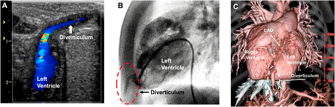
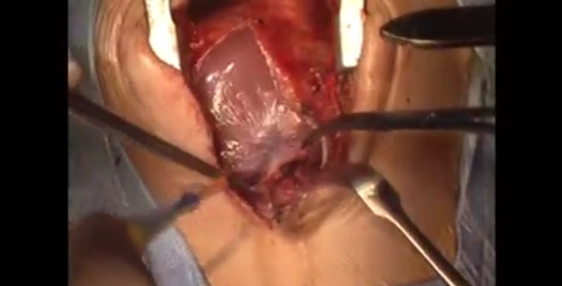
Surgical Repair of Congenital Left Ventricular Diverticulum

1Departments of Cardiovascular Surgery, Japan Community Health Care Organization (JCHO), Kyushu Hospital ◇ Fukuoka, Japan

2Pediatric Cardiology, Japan Community Health Care Organization (JCHO), Kyushu Hospital ◇ Fukuoka, Japan

受付日:2020年5月2日Received: May 2, 2020
受理日:2020年6月29日Accepted: June 29, 2020
発行日:2021年3月1日Published: March 1, 2021
HTMLPDFEPUB3

Key words: left ventricle; diverticulum; surgical repair

Introduction

Congenital left ventricular (LV) diverticulum is often associated with cardiac rotation disorder (dextrocardia or mesocardia) and/or intracardiac anomalies such as ventricular septal defect (VSD), atrial septal defect, and tetralogy of Fallot.1) Congenital LV diverticulum can potentially give rise to thromboembolism, arrhythmias, infection, cardiac failure, and rupture.2) Here, we report the surgical repair of a congenital LV diverticulum.

Case Report

A 4-month-old boy weighing 5.2 kg, with failure to thrive has presented with a prominent pulsatile upper anterior abdominal wall swelling, which was initially diagnosed as an umbilical anomaly. The patient was the first child of his mother, who gave birth after 38 weeks of gestation. His birth weight was 2.52 kg. The clinical examination confirmed the presence of a subcutaneous mass that showed synchronous pulsatility, consistent with a heartbeat (Video 1). Doppler ultrasonography demonstrated blood flow in and out of the pulsatile mass, which contracted in synchrony with the myocardium (Fig. 1A). Echocardiography revealed a large perimembranous outlet VSD. Angiography (Fig. 1B) and computed tomography (Fig. 1C) showed a contrast-filled evagination from the LV apex, along with cardiac rotation disorder (mesocardia), severe pulmonary hypertension, and a persistent left superior vena cava.

Journal of Pediatric Cardiology and Cardiac Surgery 5(1): 36-37 (2021)

Video 1

Journal of Pediatric Cardiology and Cardiac Surgery 5(1): 36-37 (2021)

Fig. 1 (A) Echocardiography demonstrated blood flow in and out of the pulsatile mass. The white arrow indicates the diverticulum. (B) Angiography of left ventricle shows the diverticulum (red dotted circle) as an outpouching of the left ventricular apex. (C) Computed tomographic angiography shows the diverticulum (white dotted circle) as an elongated contrasted-filled outpouching arising from the left ventricular apex. The neck of the diverticulum is near the left anterior descending coronary artery (white arrow). LAD, left anterior descending.

The exit of the diverticulum from the LV was small, and the blind end of the skin was also tiny. Therefore, we planned the resection of the LV diverticulum and patch closure of the VSD were performed concomitantly. The skin incision was carefully extended from just below the suprasternal notch beyond the xiphoid process to the pulsatile tumor. The sternum was cut from top to bottom to avoid injuring the pulsatile tumor. After pericardial incision, the diverticulum was easily accessed because of the mesocardia. The relationship between the neck of the diverticulum and the left anterior descending coronary artery was confirmed so that the coronary was not damaged. Subsequently, the neck of the diverticulum was ligated and the blind end of the diverticulum and associated abdominal skin was resected prior to cardiopulmonary bypass, as shown in Video 2. The postoperative course was uneventful. Currently, 13 years after surgery, the patient remains in good condition.

Journal of Pediatric Cardiology and Cardiac Surgery 5(1): 36-37 (2021)

Video 2

Discussion

Synchronous contractility that has been recognized as the most reproducible parameter for the diagnosis of congenital LV diverticulum.3, 4) In the present patient, echocardiography showed a pulsatile mass that contracted in synchrony with the myocardium, and LV diverticulum was diagnosed. Two categories of congenital LV diverticulum can be identified on the basis of their location: apical and non-apical. Apical congenital diverticulum has a common defect in embryologic development with midline thoracoabdominal formation. The mechanism may be result from a failure of normal fusion of the paired primitive mesoderm in combination with abnormal fusion of the cardiac loop to the yolk sac.1)

The ideal treatment for LV diverticulum remains to be determined. The prognosis for apical congenital LV diverticulum depends on the associated intracardiac malformations, but is generally good after repair. When symptomatic or when associated with other cardiac abnormalities, surgical treatment is usually recommended.1) Malakan et al.5) reported a novel classification for congenital LV outpouching, and suggested that this classification is a useful tool that aids in treatment selection and enables the prediction of prognosis.

In the present case, we successfully performed surgical repair of a LV diverticulum in a patient with a large perimembranous VSD and severe pulmonary hypertension.

謝辞Acknowledgments

We thank Mark Cleasby, PhD, from Edanz Group (www.edanzediting.com/ac) for editing a draft of this manuscript.

Conflicts of Interest

The authors have no conflicts interest to declare.

Ethical Statement

Informed consent for the publication of this case report was obtained from the patient and the patient’s guardians.

Note

The supplementary videos of this article can be viewed online.

This page was created on 2021-01-21T10:23:45.183+09:00
This page was last modified on 2021-03-23T15:46:19.000+09:00


このサイトは(株)国際文献社によって運用されています。