Journal of Pediatric Cardiology and Cardiac Surgery

Online ISSN: 2433-1783 Print ISSN: 2433-2720
Japanese Society of Pediatric Cardiology and Cardiac Surgery
Japanese Society of Pediatric Cardiology and Cardiac Surgery Academy Center, 358-5 Yamabuki-cho, Shinju-ku, Tokyo 162-0801, Japan
Journal of Pediatric Cardiology and Cardiac Surgery 5(2): 116-117 (2021)
doi:10.24509/jpccs.20-047

Images in Pediatric and Congenital Heart DiseaseImages in Pediatric and Congenital Heart Disease

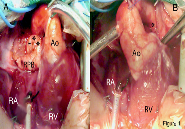
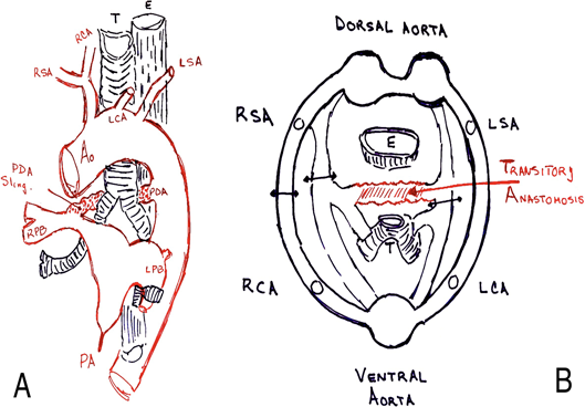
Ductus Arteriosus Sling in a Newborn with Double Inlet Left Ventricle and Pulmonary Atresia

Cardiothoracic Surgery Unit, Hospital Regional “Vicente Corral” ◇ Cuenca, Azuay, Ecuador

受付日:2020年12月17日Received: December 17, 2020
受理日:2021年3月4日Accepted: March 4, 2021
発行日:2021年7月1日Published: July 1, 2021
HTMLPDFEPUB3

Key words: ductus arteriosus; pulmonary atresia; sling

Introduction

Binet et al.1) in 1978, suggested the term “ductus arteriosus sling” describe the persistence of the ductus arteriosus which connected the right pulmonary artery to the isthmic portion of the thoracic aorta, having passed between the trachea and the esophagus. It has been considered a rare vascular ring.2, 3) We have been unable to find a published intraoperative image of this condition, and consider it worthy to report.

Clinical Course

A 7-day-old male newborn weighing 3.5 kg, referred for cyanosis. Echocardiography revealed a double inlet left ventricle and pulmonary atresia, left aortic arch, left descending aorta, and a tortuous, left patent ductus arteriosus of 4 mm in diameter. To perform an emergency right modified Blalock–Taussig shunt, a median sternotomy was used. On dissecting the superior border of the right pulmonary artery a vessel of approximately 4 mm in diameter was encountered (Fig. 1A). Diagnoses of the left pulmonary artery sling or isolated subclavian artery were ruled out. The vessel was traced between the trachea and the esophagus to the isthmic portion of the thoracic aorta, confirming the ductus arteriosus sling (Fig. 1B). We have dissected the pulmonary branch confluence and the atretic pulmonary trunk behind the aorta and confirmed the anomalous vessel was not inserting into the left pulmonary artery, but into the right. Normothermic cardiopulmonary bypass was used, ductus arteriosus was divided and a modified Blalock-Taussig shunt was performed with a 3.5 mm polytetrafluoroethylene tube. The postoperative course was without complications.

Journal of Pediatric Cardiology and Cardiac Surgery 5(2): 116-117 (2021)

Fig. 1 A) Patent ductus arteriosus (***) connecting to the superior border of the right pulmonary artery (RPA). B) Origin of the patent ductus arteriosus (*) at the aortic isthmus.

Ao, aorta; RA, right atrium; RV, right ventricle.

Discussion

As suggested by Congdon,4) in the 5-mm embryo there appears to be a plexiform communication between the right and left primary pulmonary arteries in the frontal plane and between the trachea and esophagus in the sagittal plane (Fig. 2B). This communication disappears when the embryo reaches the 8-mm stage. Therefore, the ductus arteriosus sling arising from the right pulmonary artery, passing between the esophagus and trachea and opening into the descending aorta, may represent the ductus arteriosus only in its very dorsal portion, the longest segment belong to the communication between the primary right and left pulmonary arteries (Fig. 2A).1)

Journal of Pediatric Cardiology and Cardiac Surgery 5(2): 116-117 (2021)

Fig. 2 A) Frontal view of the great vessels (drawing). The abnormal ductus is seen running behind the trachea. B) Possible embryology of the anomaly. Transverse section of 8-mm stage embryo. Transitory plexiform communication is between right and left pulmonary artery (Red arrow). Black arrows show the normal points of involution of the aortic arch.

Ao, aorta; E, esophagus; LCA, left common artery; LPB, left pulmonary branch; LSA, left subclavian artery; PA, pulmonary atresia; RCA, right common artery; RPB, right pulmonary branch; RSA, right subclavian artery; T, trachea.

謝辞Acknowledgments

The authors would like to thank Diane Alejo at the Johns Hopkins Medical Institutions for her assistance with this manuscript.

Conflicts of Interest

The authors have no conflicts of interest to declare.

引用文献References

1) Binet JP, Conso JF, Losay J, et al: Ductus arteriosus sling: Report of a newly recognized anomaly and its surgical correction. Thorax 1978; 33: 72–75

2) Backer CL, Mavroudis C: Congenital heart surgery nomenclature and database project: Vascular rings, tracheal stenosis, pectus excavatum. Ann Thorac Surg 2000; 69 Suppl: S308–S318

3) Waldo K, Kirby ML: Development of the Great Arteries, in de la Cruz MV, Markwald RR (eds): Living Morphogenesis of the Heart. 1st edition. Boston, Springer Science & Business Media, 1998, pp 187–218

4) Congdon ED: Transformation of the aortic arch system during the development of the human embryo. Contrib Embryol 1922; 14: 47–110

This page was created on 2021-06-18T10:40:02.601+09:00
This page was last modified on 2021-07-09T15:33:32.000+09:00


このサイトは(株)国際文献社によって運用されています。