Journal of Pediatric Cardiology and Cardiac Surgery

Online ISSN: 2433-1783 Print ISSN: 2433-2720
Japanese Society of Pediatric Cardiology and Cardiac Surgery
Japanese Society of Pediatric Cardiology and Cardiac Surgery Academy Center, 358-5 Yamabuki-cho, Shinju-ku, Tokyo 162-0801, Japan
Journal of Pediatric Cardiology and Cardiac Surgery 6(2): 66-69 (2022)
doi:10.24509/jpccs.21-032

Case ReportCase Report

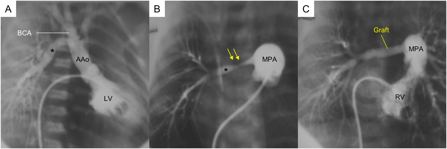
Anomalous Origin of the Right Pulmonary Artery from the Ascending Aorta with Aortic Coarctation

1Department of Cardiovascular Surgery, JCHO Kyushu Hospital ◇ Fukuoka, Japan

2Department of Pediatric Cardiology, JCHO Kyushu Hospital ◇ Fukuoka, Japan

受付日:2021年12月22日Received: December 22, 2021
受理日:2022年2月15日Accepted: February 15, 2022
発行日:2022年7月1日Published: July 1, 2022
HTMLPDFEPUB3

Two neonates were treated for a combination of a distal type of anomalous origin of the right pulmonary artery from the ascending aorta and aortic coarctation. Aortic arch reconstruction was performed using an extended aortic arch anastomosis in both cases. In the first case, the right pulmonary artery was reconstructed by a direct anastomosis behind the ascending aorta. Early reoperation for graft interposition was required to treat stenosis at the proximal right pulmonary artery due to a residual ductal tissue there. In the second case, such ductal tissues were sufficiently resected at the initial repair, and the right pulmonary artery was reconstructed using an artificial graft anterior to the ascending aorta. As aortic arch reconstruction for aortic coarctation reduces the space posterior to the ascending aorta, an appropriate route should be considered sensibly for the right pulmonary artery to be reconstructed.

Key words: anomalous origin of the right pulmonary artery; cardiac defect; coarctation of the aorta; persistent fifth aortic arch

Introduction

Anomalous origin of the right pulmonary artery from the ascending aorta (AORPA) is a rare congenital cardiac defect that has been classified into a proximal and a distal forms.1) Epidemiological studies have reported that AORPA usually occurs in isolation. The malformation can coexist with aortic arch hypoplasia and occasionally with arch obstruction.2) We encountered two neonates with AORPA of the distal type and coarctation of the aorta (CoA).

Case Report

Case 1

A boy weighing 3.0 kg, with AORPA of the distal form, aortic arch hypoplasia, atrial septal defect, and patent ductus arteriosus (PDA), underwent primary surgical repair at 7 days old. The results of a PDA occlusion test revealed a significantly reduced descending aortic pressure, which was consistent with CoA. After the CoA repair with an extended aortic arch anastomosis, the right pulmonary artery (RPA) arising immediately proximal to the bracheocephalic artery (Fig. 1A) was transected at its origin and reconstructed with direct anastomosis to the main pulmonary arterial trunk (MPA) behind the ascending aorta (AAo). Postoperative MPA angiography illustrated a diffuse stenosis of the proximal RPA (Fig. 1B). Subsequently, the patient underwent the second operation for excision of the stenotic lesion and graft interposition using a 5-mm expanded polytetrafluoroethylene graft (Gore-Tex; W.L. Gore and Associates, Flagstaff, AZ, USA) behind the AAo at 1 month following the initial operation. The histological specimen of the excised vessel proved to have a remnant of a PDA tissue. Postoperative cardiac catheterization showed a large RPA that had been reconstructed without compression from the AAo, and the ratio of pulmonary to systemic arterial pressures decreased from 1.0 to 0.31 (Fig. 1C). Nine years later, the artificial graft was upsized to a 14-mm expanded polytetrafluoroethylene graft. This patient is currently 25 years old and remains healthy.

Journal of Pediatric Cardiology and Cardiac Surgery 6(2): 66-69 (2022)

Fig. 1 Pre- and postoperative angiography in Case 1

A: Preoperative left ventriculogram.B: Pulmonary arterial angiogram after the initial repair. C: Rght ventriculogram after revision of the right pulmonary artery. AAo, ascending aorta; BCA, bracheocephalic artery; LV, left ventricle; MPA, main pulmonary arterial trunk; RV, right ventricle. Black asterisk: right pulmonary artery; yellow arrows: stenosis.

Case 2

A boy weighing 2.5 kg was transferred to our hospital on the first day of life with desaturation and diagnosed as having distal AORPA, CoA, small PDA, and ventricular septal defect (VSD) on echocardiography. Preoperative computed tomography revealed that the RPA originated from the distal portion of the AAo immediately proximal to the bracheocephalic artery with severe stenosis at its origin (Fig. 2A). Severe CoA was present and the transverse arch was hypoplastic (Fig. 2B). Pulmonary overcirculation started at 1 day old, and progressed gradually. The patient was managed without prostaglandin administration until 5 days old when the small PDA was closed and a minor blood pressure difference (approximately 20 mmHg) was demonstrated between the upper and the lower limbs. Primary surgical repair was performed at 16 days old. After repair of the CoA using an extended aortic arch anastomosis, the proximal RPA including the stenotic tissue (Fig. 2C) was sufficiently resected. The right and the left pulmonary arteries were well mobilized up to the hilar branches; still, tension-free reconstruction by direct anastomosis was not feasible between the distal RPA and the MPA because of their distance. The RPA was eventually reconstructed using a 6-mm expanded polytetrafluoroethylene graft in the way in which the tube was coursing anterior to the AAo and curving upwards to avoid compression from the sternum (Fig. 2D, E). Histological investigations using hematoxylin-eosin staining showed mucopolysaccharides accumulated beneath the elastic layer, which were identified as a PDA tissue (Fig. 2F). On echocardiography, the peak pressure gradient estimated by tricuspid valve regurgitation had decreased from 67 to 28 mmHg. No signs of RPA stenosis were observed 1 year postoperatively.

Journal of Pediatric Cardiology and Cardiac Surgery 6(2): 66-69 (2022)

Fig. 2 Pre- and postoperative investigations and intraoperative photos in Case 2

A: Anterior view of preoperative 3-dimensional CT. B: Posterior view of preoperative 3-dimensional CT. C: An intraoperative photo prior to reconstruction of the right pulmonary artery. D: An intraoperative photo after reconstruction of the right pulmonary artery. E: Anterior view of postoperative 3-dimensional CT. F: Histological findings of surgical specimen from the proximal right pulmonary artery. AAo, ascending aorta; BCA, bracheocephalic artery; CoA, coarctation of the aorta; CT, computed tomography; MPA, main pulmonary arterial trunk; RV, right ventricle; SVC, superior vena cava. Black asterisk: right pulmonary artery; yellow arrows: stenosis, black arrow heads: mucopolysaccharidosis.

Discussion

Embryologically, distal AORPA is caused by a persistent right fifth arch connecting to the sixth arch to form the RPA. In particular, a finding that the RPA originates just proximal to the origin of the bracheocephalic artery strongly suggests this embryological explanation and could lead to severe stenosis across the proximal RPA.2, 3) Organogenesis of the aortic arch is achieved not only through genetically programmed signals but also through hemodynamics-induced shear stress.4) In AORPA, a part of blood from the left ventricle to the AAo flows into an aberrant RPA, which resulted in decreased blood flow through the transverse arch. Concurrently, blood flow from the right ventricle into the descending aorta through the PDA increases due to the absence of the RPA. These hemodynamic flow variants due to AORPA explain the hypoplastic aortic arch with a large AAo (Fig. 3).5) In case 1, a large atrial septal defect caused an unrestricted inflow of highly oxygen-saturated blood from the inferior vena cava through the AAo into the RPA, allowing a larger amount of RPA blood flow than normal even in the fetal stage. In case 2, a left-to-right intraventricular shunt occurred due to VSD. These factors were considered to enhance the abnormal flow distribution described above, resulting in various degrees of aortic arch hypoplasia.

Journal of Pediatric Cardiology and Cardiac Surgery 6(2): 66-69 (2022)

Fig. 3 Hemodynamic schema of aortic arch formation

A: Normal. B: In AORPA of the distal form. Ao, aorta; AORPA, anomalous origin of the right pulmonary artery from the ascending aorta; DAo, descending aorta; III, IV, V, VI, aortic arches; LSCA, left subclavian artery; PA, pulmonary artery. Red arrows: blood from the left ventricle; blue arrows: blood from the right ventricle.

When RPA is reconstructed in a distal AORPA, sufficient tissue resection is crucial from the central portion of the RPA, because the wall could be comprised of the PDA tissue.2, 6) This circumstance renders direct anastomosis between the RPA and the AAo difficult. The RPA can be reconstructed via a route behind the AAo ideally using autologous tissue, such as an aortic or a pulmonary arterial flap.7, 8) In our two cases, extended aortic arch anastomosis was performed for the hypoplastic aortic arch. Arch repair using an autologous pericardial patch could have been chosen to create a retro-aortic space; however, it is also ideal to reconstruct the aortic arch without any supplemental materials. Reconstruction of the RPA interposing an artificial graft was, therefore, acceptable for repair of distal AORPA with CoA.9) In such circumstances, the route of the RPA graft should be carefully chosen according to the space available behind the reconstructed aorta. If the AAo shifted posteriorly due to the arch reconstruction and the RPA route was chosen behind the AAo, the proximal RPA would be more susceptible to compression from the AAo. In contrast, when the RPA courses anteriorly to the AAo, there is a sufficient space created in front of the AAo, accommodating an artificial graft of a sufficient size without compression from the sternum. Although graft replacement is inevitable in the future, RPA reconstruction by means of graft interposition for repair of distal AORPA with CoA should be one of the procedures of choice.

Financial Support

None.

Conflicts of Interest

None.

Ethical Standards

Informed consent was obtained from the patient or the patient’s guardians.

This page was created on 2022-06-02T14:04:49.508+09:00
This page was last modified on 2022-08-03T17:00:35.000+09:00


このサイトは(株)国際文献社によって運用されています。