Journal of Pediatric Cardiology and Cardiac Surgery

Online ISSN: 2433-1783 Print ISSN: 2433-2720
Japanese Society of Pediatric Cardiology and Cardiac Surgery
Japanese Society of Pediatric Cardiology and Cardiac Surgery Academy Center, 358-5 Yamabuki-cho, Shinju-ku, Tokyo 162-0801, Japan
Journal of Pediatric Cardiology and Cardiac Surgery 7(1): 46-48 (2023)
doi:10.24509/jpccs.22-005

Images in Pediatric and Congenital Heart DiseaseImages in Pediatric and Congenital Heart Disease

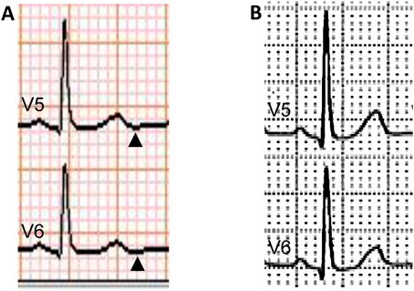
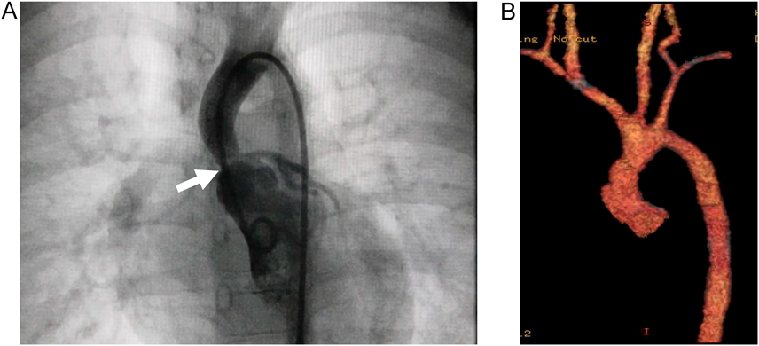
Negative U-Wave in Williams Syndrome with Supravalvar Aortic Stenosis

Department of Pediatrics, Hirosaki General Medical Center ◇ Aomori, Japan

受付日:2022年3月15日Received: March 15, 2022
受理日:2022年6月13日Accepted: June 13, 2022
発行日:2023年3月31日Published: March 31, 2023
HTMLPDFEPUB3

Key words: negative U-wave; Williams syndrome; supravalvar aortic stenosis

Introduction

Negative U-wave (NU), excluding on the aVR lead in a standard 12-lead electrocardiogram (ECG), is considered abnormal in patients with underlying cardiovascular diseases. It is recognized as a sensitive marker of myocardial impairment, which indicates the presence of ventricular pressure and volume overload or myocardial ischemia.1) Most of pediatric cardiologists are, disappointingly, less interested in NU compared with cardiologists in adult cardiovascular medicine. Presented herein is a 4-year-old girl with Williams syndrome having significant supravalvar aortic stenosis (SVAS), in whom preoperative ECG showed NU in left-sided precordial leads suggesting pressure overload to the left ventricle (LV).

Case

This patient was first diagnosed with a heart murmur at the age of 6 months. Echocardiography showed mild SVAS and mild peripheral pulmonary stenosis. She was suspected of Williams syndrome on the basis of intellectual disability, characteristic appearance, and concomitant cardiac defects at the age of 3 years. The chromosomal analysis confirmed the disorder with 7q11.23 microdeletion by fluorescence in situ hybridization. Pressure gradient of 50 mmHg was observed across SVAS on continuous-wave Doppler (CW) at the age of 4 years. LV end-diastolic septal thickness, LV end-diastolic posterior wall thickness, and LV end-diastolic diameter were 4.6 mm with z score −1.15*, 5.5 mm with z-score 1.06*, and 25.5 mm with z-score −3.12*, respectively, indicating no LV wall hypertrophy or enlargement (*according to a web site (https://zscore.chboston.org), based on body surface area with 0.57 m2; for the LV end-diastolic posterior wall, the LV end-diastolic free wall thickness on the site was applied). LV systolic function was normal. LV diastolic function, including the early to late diastolic mitral flow velocity ratio, was not evaluated. ECG did not meet the criteria for electrocardiographic LV hypertrophy; RV5=1.7 mV, RV6=1.4 mV, SV1+RV5=3.5 mV, and SV1+RV6=3.2 mV.2) NU was observed in V5 and V6 leads (Fig. 1A). Pressure study on cardiac catheterization revealed pressure difference of 50 mmHg between the LV and the ascending aorta. The ascending aorta was narrow above the Valsalva sinus on LV-graphy (Fig. 2A). After repair of SVAS (Fig. 2B), pressure gradient estimated by CW improved to 25 mmHg. At 5 months after surgery, NU in V5 and V6 leads disappeared (Fig. 1B).

Journal of Pediatric Cardiology and Cardiac Surgery 7(1): 46-48 (2023)

Fig. 1 Electrically magnified electrocardiogram (ECG)

(A) Preoperative ECG. Negative U-wave is observed in V5 and V6 leads (arrowheads). (B) Postoperative ECG. Negative U-wave vanished.

Journal of Pediatric Cardiology and Cardiac Surgery 7(1): 46-48 (2023)

Fig. 2 Radiographic images

(A) Preoperative left ventriculography. The ascending aorta is narrow above the Valsalva sinus (arrow). (B) Postoperative enhanced computed tomography. The ascending aorta appears less obstructed morphologically.

Discussion

According to the phasic relationship to positive U-wave deflection, NU is classified as initial or terminal U inversion3); the former is associated with ventricular pressure overload, such as hypertension, and is to proceed to positive U wave deflection. The latter is following positive U wave deflection, associated with myocardial ischemia, such as angina pectoris. In our patient, the observed NU in the preoperative ECG was considered as initial U inversion, suggesting significant LV pressure overload induced by SVAS although the ECG did not meet the LV hypertrophy criteria. The reason why positive U wave deflection was not observed following this initial U inversion could be that deflection was extremely small, even though it could have been magnified.

Miwa4) reported that NU disappeared according to improvement of blood pressure with antihypertensive therapy in hypertensive patients with initial U inversion, and LV diastolic dysfunction on echocardiography before treatment was normalized in the post-treatment term. Therefore, they hypothesized that initial U inversion is caused by action potential gradient formation between the endocardium and epicardium, produced by stretching induced after depolarization in the ventricular apical area with impaired early LV relaxation. LV diastolic dysfunction was not evaluated in our patient; nonetheless, NU transition seen in V5 and V6 leads supported the hypothesis of Miwa et al.

U wave deflection is commonly tiny and difficult to detect with pulse rate of 95/min or higher.5) Accordingly, it is apt to be overlooked in young children with relatively fast heart rate compared with in adults. NU may be more easily magnified and detected using a personal computer or other electronic media. Using such modern techniques, NU presenting with the initial U wave inversion may reflect early ventricular pressure or volume overload before ventricular hypertrophy becomes evident on ECG. Furthermore, in our patient, no clinical symptoms such as angina nor coronary arterial abnormalities were observed on aortography; even so, the essence of NU with the terminal U inversion could have been noted concomitantly, considering myocardial ischemia due to coronary arterial impairment which is known to be a serious complication often occurring in Williams syndrome with SVAS.

In conclusion, pediatric cardiologists in charge of treating congenital heart disease should take a keen interest in NU for electrocardiographic interpretation.

謝辞Acknowledgments

We thank the pediatric cardiology and cardiovascular surgery staff of Hirosaki University Hospital (in Aomori, Japan) for performing the cardiac catheterization and repair in this case.

Conflicts of Interest

The authors declare no conflict of interest.

This page was created on 2022-12-02T14:26:32.946+09:00
This page was last modified on 2023-03-23T16:32:46.000+09:00


このサイトは(株)国際文献社によって運用されています。