Journal of Pediatric Cardiology and Cardiac Surgery

Online ISSN: 2433-1783 Print ISSN: 2433-2720
Japanese Society of Pediatric Cardiology and Cardiac Surgery
Japanese Society of Pediatric Cardiology and Cardiac Surgery Academy Center, 358-5 Yamabuki-cho, Shinju-ku, Tokyo 162-0801, Japan
Journal of Pediatric Cardiology and Cardiac Surgery 7(1): 52-53 (2023)
doi:10.24509/jpccs.22-010

Images in Pediatric and Congenital Heart DiseaseImages in Pediatric and Congenital Heart Disease

Four-Dimensional Flow Magnetic Resonance Imaging Can Visualize a Disturbed Pattern of Blood Flow in a Patient Without a Significant Pressure Gradient after Surgical Repair of Aortic Coarctation

1Department of Pediatrics, Asahikawa Medical University ◇ Hokkaido, Japan

2Section of Radiological Technology, Department of Medical Technology, Asahikawa Medical University Hospital ◇ Hokkaido, Japan

受付日:2022年5月11日Received: May 11, 2022
受理日:2022年9月8日Accepted: September 8, 2022
発行日:2023年3月31日Published: March 31, 2023
HTMLPDFEPUB3

Key words: aortic coarctation; 4D flow MRI; helical flow; wall shear stress; energy loss

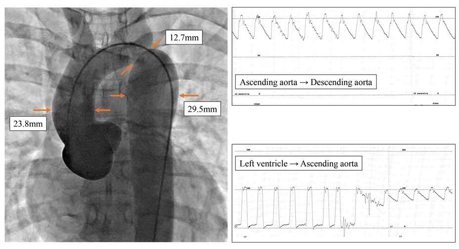
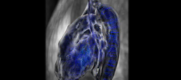
An 18-year-old male was admitted to hospital for postoperative evaluation, who had possessed aortic coarctation initially and undergone extended aortic arch anastomosis at the age of 16 days. His aortic valve is bicuspid. Blood pressure did not differ between the upper and lower extremities. Cardiac catheterization showed no pull-back pressure gradient across the aortic valve or the anastomosis, although the anastomotic site appeared stenotic (a diameter 12.7 mm compared to 23.8 mm at the ascending aorta) (Supplementary Fig. S1). The descending aorta was dilated up to 29.5 mm. Four-dimensional flow magnetic resonance imaging (4D flow MRI) showed marked helical flow in the descending thoracic aorta from systole to diastole (Fig. 1A; Supplementary Movie S1). Averaged wall shear stress and peak energy loss were 0.27 Pa and 1.21 mW, respectively, at the anastomosis; these values were markedly raised compared with the 0.10 Pa and 0.55 mW in the ascending aorta and 0.15 Pa and 0.43 mW in the descending aorta (Fig. 1B, C). The workstation Cvi42 (Circle, Cardiovascular Imaging, Calgary, Canada) was used for this analysis.

Journal of Pediatric Cardiology and Cardiac Surgery 7(1): 52-53 (2023)

Fig. 1 (A) Four-dimensional flow magnetic resonance imaging showed marked helical flow in the descending thoracic aorta. (B, C) Averaged wall shear stress and peak energy loss at the anastomotic site were elevated compared to those in the ascending and the descending aorta.

4D flow MRI can visualize a pressure gradient across a stenosed anastomosis.1, 2) It remains unclear, in contrast, whether the pattern of blood flow is altered beyond the morphologically obstructed site but no significant pressure gradient. Disturbed patterns of blood flow are caused by changes in aortic geometry after surgical repair of aortic coarctation, especially when the aorta forms the shape of a “gothic” type in which helical flow tends to occur in the descending aorta, or a “crenel” type in which helical flow occurs in 40% of cases.1, 3) In our patient, the configuration of the reconstructed aorta was of a “crenel” type, and helical flow probably occurred because of this feature. The presence of helical flow has been reported to affect hypertension and aortic dilatation; this consequence is likely associated with elevated wall shear stress in the area where helical flow occurs.1) When evaluating energy loss, it should be noted that its actual values are not reproducible and the values derived from different examinations cannot be simply compared. This is because multiple spin velocities of turbulent flow within a voxel causes signal loss on phase contrast MRI.4) The follow-up of patients after repair of aortic coarctation often focuses on whether a restenosis has progressed or not. Unless there is a significant obstruction at the anastomosis, those patients would not undergo extensive examinations. Even in young patients without a significant restenosis, however, aortic blood flow may be abnormal.

4D flow MRI is an important tool to identify such abnormal blood flow and to provide an early warning of potential impediments in patients undergoing aortic reconstruction.

Note

Supplementary Figure and Movie are provided online for this article.

Journal of Pediatric Cardiology and Cardiac Surgery 7(1): 52-53 (2023)

Supplementary Fig. S1 Aortic angiography shows that the previous anastomosis has become stenotic. Cardiac catheterization did not record a significant pull-back pressure gradient across the aortic valve or the anastomosis.

Journal of Pediatric Cardiology and Cardiac Surgery 7(1): 52-53 (2023)

Supplementary Movie

Conflicts of Interest

The authors have no conflicts of interest to declare.

IRB Information

Asahikawa Medical University Ethics Committee, 19250.

This page was created on 2022-12-07T15:52:33.298+09:00
This page was last modified on 2023-03-13T13:52:03.000+09:00


このサイトは(株)国際文献社によって運用されています。