Journal of Pediatric Cardiology and Cardiac Surgery

Online ISSN: 2433-1783 Print ISSN: 2433-2720
Japanese Society of Pediatric Cardiology and Cardiac Surgery
Japanese Society of Pediatric Cardiology and Cardiac Surgery Academy Center, 358-5 Yamabuki-cho, Shinju-ku, Tokyo 162-0801, Japan
Journal of Pediatric Cardiology and Cardiac Surgery 8(2): 77-81 (2024)
doi:10.24509/jpccs.23-009

Case ReportCase Report

Reimplantation for Anomalous Aortic Origin of Right Coronary Artery in the Elderly

Department of Cardiovascular Surgery, Kinan Hospital ◇ Wakayama, Japan

受付日:2023年5月10日Received: May 10, 2023
受理日:2023年10月12日Accepted: October 12, 2023
発行日:2024年7月1日Published: July 1, 2024
HTMLPDFEPUB3

Anomalous aortic origin of the right coronary artery (AAORCA) is a rare congenital anomaly. It is even rarer in the elderly, and there is ongoing controversy regarding techniques for surgical intervention. We describe a case of AAORCA that was successfully treated using a reimplantation technique. A 65-year-old female with a history of exercise-related angina was referred to our hospital. Enhanced computed tomography (CT) revealed AAORCA. Ischemia was detected on electrocardiogram exercise testing, although myocardial perfusion scintigraphy had failed to show it; thus, surgical intervention was recommended. We applied a reimplantation technique, because the right coronary artery (RCA) was not hypoplastic but its proximal course was interarterial with a slit-like ostium. We transected the RCA beyond the intramural portion and closed the residual stump proximally. An aortic wall was punched out higher in the right facing sinus of Valsalva to create a neo-ostium. The RCA was spatulated on the inferior side and anastomosed to the neo-ostium. The patient’s postoperative course was uneventful. Postoperative enhanced CT showed neither stenosis nor kinking of the reimplanted vessel. The patient has been asymptomatic for more than 2 years since the operation. The reimplantation technique was useful for AAORCA with a slit-like ostium and an interarterial course. Its medium- to long-term outcomes are reported to be satisfactory. Therefore, the reimplantation technique could easily be applied to similar cases.

Key words: anomalous aortic origin of right coronary artery; coronary artery anomaly; reimplantation technique; unroofing technique; coronary artery bypass grafting

Introduction

Anomalous aortic origin of the right coronary artery (AAORCA) is a rare congenital anomaly that may cause sudden cardiac death, especially in young athletes.1) It is quite rare to find the malformation in the elderly. The 2018 Adult Congenital Heart Disease (ACHD) Guideline2) states that surgery is recommended for AAORCA in patients with symptoms, or diagnostic evidence consistent with coronary ischemia, attributable to the anomalous coronary artery. As for surgical intervention, there is ongoing controversy over the techniques, especially in the elderly. Here we describe a case of an elderly patient whose evaluation of episodic chest pain revealed AAORCA. Her right coronary artery (RCA) had an interarterial course with a slit-like ostium and it was successfully treated using a reimplantation technique.

Case Report

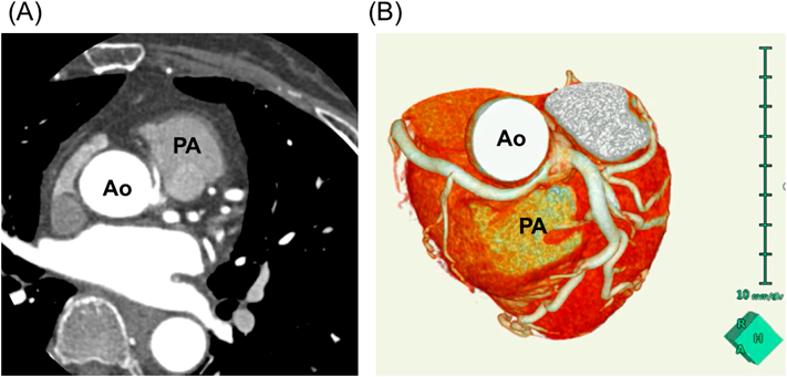
A 65-year-old woman with a history of exercise-related angina was referred to our hospital. She had no history of syncope or ventricular arrhythmia, and no coronary risk factors. The electrocardiogram at rest was within normal limits, and echocardiogram showed no left ventricular asynergy. However, laboratory data indicated elevated troponin I (74.5 pg/mL). She underwent coronary angiography, which revealed AAORCA. Coronary 3D computed tomography (CT) showed the RCA departing independently from the left coronary sinus of Valsalva and passing between the ascending aorta and the pulmonary trunk (the so-called interarterial course) (Fig. 1A, B).

Journal of Pediatric Cardiology and Cardiac Surgery 8(2): 77-81 (2024)

Fig. 1 Enhanced computed tomography before the operation

(A) Axial view (viewing from below) showing that the right coronary artery (RCA) arises from the left coronary sinus of Valsalva and passes between the aorta (Ao) and the pulmonary artery (PA). (B) Cranial view (from above) of 3D computed tomography showing that the course of the RCA is interarterial and that the interarterial portion is flattened. Ao, aorta; PA, pulmonary artery.

Myocardial perfusion scintigraphy failed to exhibit ischemia; nonetheless, electrocardiogram exercise testing revealed ST depression in leads II, III, and aVF. Thus, we recommended surgical intervention based on the guideline.2)

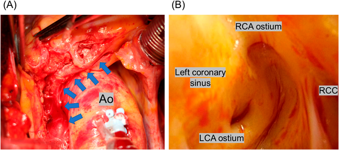
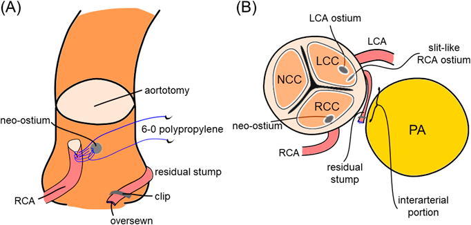
Through a median sternotomy, the AAORCA was identified (Fig. 2A); using a harmonic scalpel, we dissected the proximal portion of the RCA just distal to the intramural portion for 2 cm, sparing the proximal branches. The superior aspect of the RCA was marked to prevent distortion. Cardiopulmonary bypass was established by cannulation to the ascending aorta and the right atrium. The left ventricular venting tube was inserted via the right upper pulmonary vein. The initial cardioplegia was infused antegradely from the aortic root. A transverse aortotomy was performed 15 mm above the sino-tubular junction. We confirmed that both the left coronary artery and the RCA originated independently from the left coronary sinus of Valsalva, and that the RCA ostium was slit-like (Fig. 2B). We transected the RCA adjacent to the intramural portion. The residual proximal stump was 10 mm long. The stump was clipped, and the amputated end was oversewn with 7-0 polypropylene. The wall of the very proximal ascending aorta, just above the sino-tubular junction on the right facing sinus aspect, was punched out to create a neo-ostium. The RCA was spatulated for 4 mm on the inferior side and was anastomosed to the neo-ostium using running 6-0 polypropylene beginning at the heel. The aortotomy was closed in two layers. After infusion of warm cardioplegia, the aorta was unclamped. Electrocardiogram showed normal sinus rhythm with no significant ST-T change. The RCA flow was measured intraoperatively as 54 mL/min using doppler ultrasound. A schematic illustration of the surgery is shown in Fig. 3A, B.

Journal of Pediatric Cardiology and Cardiac Surgery 8(2): 77-81 (2024)

Fig. 2 Intraoperative images

(A) The anomalous origin and the course of the right coronary artery (RCA). Arrows indicate the anomalous aortic origin of the right coronary artery (AAORCA). The dashed circle shows the precise site of a neo-ostium. (B) Ostia of the left and the right coronary arteries (LCA and RCA) originating from the left facing sinus of Valsalva. The ostium of the RCA was slit-like. Ao, aorta; LCA, left coronary artery; LCC, left coronary cusp; RCA, right coronary artery; RCC, right coronary cusp.

Journal of Pediatric Cardiology and Cardiac Surgery 8(2): 77-81 (2024)

Fig. 3 Schematic illustration of the operation

LCA, left coronary artery; LCC, left coronary cusp; NCC, non-coronary cusp; PA, pulmonary artery; RCC, right coronary cusp.

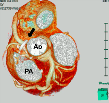
The patient’s postoperative course was uneventful. She started to take aspirin on postoperative day 1. The postoperative coronary 3D CT taken before discharge showed neither stenosis nor kinking of the reconstructed RCA (Fig. 4), and the patient was discharged on postoperative day 13. She has been asymptomatic for more than 2 years since the operation.

Journal of Pediatric Cardiology and Cardiac Surgery 8(2): 77-81 (2024)

Fig. 4 Enhanced computed tomography (CT) after the operation

Cranial view (from above) of 3D CT showing that the right coronary artery (RCA) has been reimplanted to the neo-ostium. The RCA had neither stenosis nor kinking. The arrow indicates a coronary locator.

Discussion

Anomalous aortic origin of the coronary artery (AAOCA) was first described by White and Edwards in 1948.3) The prevalence of AAORCA is reported to be 0.107% to 0.2%.4, 5) The pathomechanism of high-risk anatomic features during physical exercise includes a) kinking of the anomalous vessel with an acute-angled take-off, b) lateral compression caused by the aorta onto the intramural segment, c) intermittent closure of a slit-like orifice, and d) compression onto the interarterial portion by the aorta and the pulmonary artery.6) In our patient, c) and d) were the main causes of ischemia.

The 2018 ACHD Guideline2) states that, for anomalous aortic origin of the left coronary artery from the right sinus, surgery is reasonable even in the absence of symptoms or ischemia (class IIa). For AAORCA, on the other hand, surgery is recommended only in the presence of symptoms or diagnostic evidence consistent with coronary ischemia attributable to the anomalous coronary artery (class I). Otherwise, surgery is reasonable in the setting of ventricular arrhythmias (class IIa). In our patient, we were initially unable to detect ischemia using myocardial perfusion scintigraphy. But the cardiologist decided to check using electrocardiogram exercise testing, despite its risk of ventricular arrhythmia, after obtaining informed consent. This was because the patient’s chief complaint, exercise-related angina, seemed to be strongly related to this anomaly.

Several techniques may be used for surgical repair of AAORCA, including coronary artery bypass grafting (CABG), unroofing, reimplantation, and pulmonary arterial translocation.7, 8) CABG is a well-established technique that can be done without cardiopulmonary bypass. With this procedure, however, there is a concern regarding competitive flow at rest, and accordingly long-term patency. A short bypass using the great saphenous vein (e.g., aorta–great saphenous vein–RCA in this case) may have better patency. Moreover, myocardial infarction would not occur immediately, even if the bypass became non-patent.

The unroofing technique is the surgical option most widely used for intramural AAOCA.9) As discussed above, however, the probable pathomechanisms of ischemia in our patient were intermittent closure of the slit-like orifice and compression onto the interarterial portion by the aorta and the pulmonary artery. The unroofing technique should be effective for the type of a slit-like orifice, but would not be for the interarterial circumstance.

In contrast, reimplantation techniques can be applied to either intramural or interarterial AAOCA, and they seem to have good medium- to long-term results regarding symptom-free survival.10–12) Law et al. reported 16 cases of AAORCA repaired by reimplantation.10) Surgical reimplantation was successful in 15 patients (94%) without operative mortality, and all patients were symptom-free after surgery. Cubero et al. reported 13 cases of AAORCA11); all patients are asymptomatic and have returned to exercise without limitations. Bonilla-Ramirez et al. reported 16 cases of AAOCA12); one patient underwent CABG after reimplantation because of persistent postoperative ischemic changes, while 15 patients (94%) were released to unrestricted exercise activities. Also, a recent report has shown that the commissural manipulation associated with unroofing technique may leads to the future aortic insufficiency,13) which may support the choice of the reimplantation technique in the present patient.

Because reimplantation has a potential risk of stretching, kinking, distortion, or stenosis at the anastomosis site, it is difficult to use in patients with a small coronary artery; for example, pediatric patients or patients with a hypoplastic RCA. In our case, we chose reimplantation because the RCA was not hypoplastic but was interarterial with a slit-like ostium. The patient was 65 years old. Thus, we expected a good long-term result.

Pulmonary arterial translocation may be an alternative technique in selected cases with an anomalous course of the coronary artery as a part of complex coronary arterial anatomy; in such an occasion, the risk of coronary reimplantation may outweigh the anticipated benefits. In particular, surgical options may be limited in younger patients.14)

There are some knacks related to the reimplantation techniques, such as 1) mobilization of the RCA sparing the proximal branches of the RCA, 2) reimplantation of the RCA higher in the right facing sinus or at the very proximal ascending aorta, and 3) confirmation of correct positioning of the neo-ostium about the aortic valve commissures.15) We employed the reimplantation technique for the first time; still, the outcome seemed excellent based on adequate knowledge for these approaches obtained prior to surgery. In this sense, the reimplantation technique can be reproducibly applied to similar cases.

In conclusion, we report a case of reimplantation of the RCA in an elderly patient with AAORCA. The surgical technique used is to be selected according to the patient’s age, morphology of the coronary arteries, and other comorbidities. The reimplantation technique is useful and would be of a surgical option of choice for similar cases.

Conflicts of Interest

The authors declared no potential conflicts of interest with respect to the research, authorship, and/or publication of this article.

Ethical Considerations

Informed consent was obtained from the patient to publish a case report.

Author Contribution

Kazuki Tanimoto drafted the original manuscript. Kazuki Tanimoto, Masayuki Sakaki, Tatsuya Oyama and Hiroki Mizoguchi treated the patient. Kazuki Tanimoto and Tatsuya Oyama contributed to the acquisition of clinical data and imaging. Nobuo Sakagoshi supervised the conduct of this case. All authors reviewed the manuscript draft and revised. All authors approved the final version of the manuscript to be published.

引用文献References

1) Cheitlin MD, De Castro CM, McAllister HA: Sudden death as the complication of anomalous left coronary origin from the anterior sinus of Valsalva: A not-so-minor congenital anomaly. Circulation 1974; 50: 780–787

2) Stout KK, Daniels CJ, Aboulhosn JA, et al: 2018 AHA/ACC guideline for the management of adults with congenital heart disease: Executive summary: A report of the American College of Cardiology/American Heart Association Task Force on Clinical Practice Guidelines. J Am Coll Cardiol 2019; 73: 1494–1563

3) White NK, Edwards JE: Anomalies of the coronary arteries: Report of four cases. Arch Pathol (Chic) 1948; 45: 766–771

4) Yamanaka O, Hobbs RE: Coronary artery anomalies in 126,595 patients undergoing coronary arteriography. Cathet Cardiovasc Diagn 1990; 21: 28–40

5) Cheezum MK, Liberthson RR, Shah NR, et al: Anomalous aortic origin of a coronary artery from the inappropriate sinus of Valsalva. J Am Coll Cardiol 2017; 69: 1592–1608

6) Gräni C, Kaufmann PA, Windecker S, et al: Diagnosis and management of anomalous coronary arteries with a malignant course. Interv Cardiol 2019; 14: 83–88

7) Tavaf-Motamen H, Bannister SP, Corcoran PC, et al: Repair of anomalous origin of right coronary artery from the left sinus of Valsalva. Ann Thorac Surg 2008; 85: 2135–2136

8) Gulati R, Reddy VM, Culbertson C, et al: Surgical management of coronary artery arising from the wrong coronary sinus, using standard novel approaches. J Thorac Cardiovasc Surg 2007; 134: 1171–1178

9) Buratto E, Konstantinov IE: Current surgical management of anomalous aortic origin of a coronary artery. J Thorac Cardiovasc Surg 2021; 161: 206–212

10) Law T, Dunne B, Stamp N, et al: Surgical results and outcomes after reimplantation for the management of anomalous aortic origin of the right coronary artery. Ann Thorac Surg 2016; 102: 192–199

11) Cubero A, Crespo A, Hamzeh G, et al: Anomalous origin of right coronary artery from left coronary sinus: 13 cases treated with the reimplantation technique. World J Pediatr Congenit Heart Surg 2017; 8: 315–320

12) Bonilla-Ramirez C, Molossi S, Sachdeva S, et al: Outcomes in anomalous aortic origin of a coronary artery after surgical reimplantation. J Thorac Cardiovasc Surg 2021; 162: 1191–1199

13) Jegatheeswaran A, Devlin PJ, Williams WG, et al: Outcomes after anomalous aortic origin of a coronary artery repair: A Congenital Heart Surgeons’ Society Study. J Thorac Cardiovasc Surg 2020; 160: 757–771

14) Rodefeld MD, Culbertson CB, Rosenfeld HM, et al: Pulmonary artery translocation: A surgical option for complex anomalous coronary artery anatomy. Ann Thorac Surg 2001; 72: 2150–2152

15) Grau JB, Rahmouni K, Castillo J, et al: Reimplantation for anomalous right coronary artery. JTCVS Tech 2021; 7: 226–228

This page was created on 2024-06-13T08:56:31.539+09:00
This page was last modified on 2024-07-19T15:09:13.000+09:00


このサイトは(株)国際文献社によって運用されています。