11.2. CQ 1: Is Color Doppler Effective for Screening/Diagnosing FHD?
発行日:2023年12月1日Published: December 1, 2023
© 2023 特定非営利活動法人日本小児循環器学会© 2023 Japanese Society of Pediatric Cardiology and Cardiac Surgery
The previous fetal echocardiography guidelines stated that “color Doppler was not essential for the level I fetal heart screening, but its use may improve the screening rate.” It has been reported nowadays that color Doppler imaging is useful for detecting heart diseases and visualizing intracardiac structures. The inclusion of color Doppler imaging as an essential element of the level I fetal heart screening remains controversial. Therefore, the current findings have been summarized regarding the effectiveness of color Doppler imaging for screening and diagnosing FHD.
The improvement of the detection rate/diagnosis rate of FHD with the additional use of color Doppler imaging should be determined. No previous study has reported exactly these outcomes. Several reports have mentioned that color Doppler imaging may be effective in terms of other various outcomes. These broadly included the detection rate of heart disease and the successful visualization rate of intracardiac structures or basic cross-sections. Therefore, the studies with those two outcomes set were evaluated respectively.
The primary extraction included a total of 456 reports regarding the effectiveness of color Doppler imaging for screening/diagnosing FHD. Of these, 36 were selected during the secondary extraction, and 14 fulfilled the final criteria: two case-control studies,1, 2) one randomized control trial,3) six cross-sectional studies,4–9) and five other types of reports.10–14) Of these 14articles, seven focused on the first trimester. The current guidelines recommend that fetal heart screening should be performed twice, once between 18 and 20 weeks’ gestation and the second procedure at approximately 30 weeks’ gestation. As few reports have been published directly related to this CQ, studies that focused on fetal echocardiograms during the first trimester were also included.
A systematic review was conducted on seven observational studies that focused on the detection rate of heart disease and the successful visualization rate of intracardiac structures or basic cross-sections.

Two studies described the heart disease detection rate.2, 13) It is less than perfect to establish a rigorous evaluation due to the small amount of data. Considering the heterogeneity, the overall evidence was described as a fixed effect. The confidence interval (CI) of the odds ratio did not extend beyond 1.0. It is therefore determined that color Doppler imaging is useful for detecting heart disease (odds ratio [OR], 2.64; 95% CI, 1.13–6.18; p=0.025).
The study by Eggebø et al. reported that 35% of the detected heart diseases were by means of color Doppler imaging.13) Similarly, the report by Nadel et al.2) stated that 25% of the detected heart diseases were on color Doppler imaging. Furthermore, 75% of the heart defects detected on color Doppler imaging were pulmonary valve stenosis. The results of these studies suggested that color Doppler imaging was particularly useful for detecting malformations (such as pulmonary valve stenosis) that had not exhibited prominent abnormalities on a conventional mode of echocardiography.2)
Analyzed were 5 reports regarding the visualization rate of cardiovascular structures or basic cross-sectional images. Of the five, 3 reports focused on the visualization rate of cardiovascular structures.4, 8, 14) Two described first-trimester assessments in which the visualization rate of multiple cardiovascular structures was higher when color Doppler imaging was combined with the B mode, compared with use of the B mode alone.4, 8) The study by Dong et al. that assessed the detection rate of the pulmonary veins during 12–22 weeks’ gestation showed that the rate was higher when color Doppler imaging was utilized.14) Total anomalous pulmonary venous connection is severe CHD that usually requires treatment during the early postnatal period. The efficacy of color Doppler imaging to detect this malformation is of great significance clinically. It is known that use of conventional methods alone would produce a rather low detection rate.
The remaining 2 reports were also analyzed regarding the visualization rate of basic cross-sectional images.3, 10) Either of these studies was conducted during the first trimester. The visualization rate was higher when color Doppler imaging was used in conjunction with B mode imaging, compared to B mode imaging used in isolation.
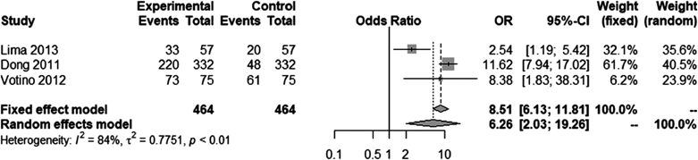
Among the 5 reports aforementioned, three3, 10, 14) were used for further assessment this time. Considering the heterogeneity, the overall evidence was described by the random effects model. The CI of the OR was wide, but it did not go over 1.0. Therefore, color Doppler imaging was useful for depicting cardiovascular structures (hazard ratio, 6.25; 95% CI, 2.03–19.26; p=0.014).
None of the five reports directly demonstrated an improved rate of diagnosing heart diseases. Even so, the ability to accurately depict the cardiovascular structure on basic cross-sectional views are naturally helpful for detecting these abnormalities. It is not unreasonable to interpret that the results of these studies reinforce effective use of color Doppler imaging. Several other studies have reported that protocols involving color Doppler imaging achieved a high detection rate for heart diseases, although these advanced approaches were not compared with the conventional method in a statistical way.6, 9, 11)
Color Doppler imaging is useful, in addition to valve diseases and anomalous pulmonary venous connection as mentioned above, for detecting other diseases. For example, ventricular septal defects are easily detected using color Doppler imaging,5) and retrograde blood flow in the aortic arch (AA) suggests the presence of coarctation of the aorta.1)
The strength of the evidence is weak due to the small number of studies. These studies mainly focused on the first trimester, which is not included in the current guidelines. Nonetheless, use of color Doppler imaging has very few risks. Many products of up-to-date ultrasonic devices are equipped with color Doppler as a part of their standard equipment. Its application would not add a significant expense. Accurate examinations would become feasible with high sensitivity and specificity. Therefore, use of color Doppler imaging for fetal heart screening/diagnosis is strongly recommended. This technique should be actively used in clinical practice, paying attention to the quality of available equipment and the skill of the examiner.
Color Doppler imaging is an ultrasound technique that visually indicates the direction of the blood flow in the cardiovascular system. Nowadays, many types of ultrasonic devices commercially available can perform color Doppler imaging.
In these guidelines, the CQ “Is color Doppler echocardiography effective for screening/diagnosing FHD?” was raised to organize the findings and to make recommendations.
The conclusions are summarized as follows:
In addition, some characteristic findings in certain diseases are not detected when the conventional ultrasound method is used in isolation. These findings would turn out to be obvious when color Doppler imaging is applied. It is recommended that color Doppler imaging should be used to screen and diagnose FHD. In practice, the technique is actively used wherever possible, taking the quality of the equipment available and the skill of the examiner into consideration.
1) Kawamura H, Inamura N, Inoue Y, et al: Is retrograde blood flow of aortic isthmus useful for the prenatal screening of coarctation of the aorta by fetal color Doppler echocardiography? A preliminary study. J Med Ultrason 2018; 45: 431–435
2) Nadel AS: Addition of color Doppler to the routine obstetric sonographic survey aids in the detection of pulmonic stenosis. Fetal Diagn Ther 2010; 28: 175–179
3) Votino C, Kacem Y, Dobrescu O, et al: Use of a high-frequency linear transducer and MTI filtered color flow mapping in the assessment of fetal heart anatomy at the routine 11 to 13+6-week scan: A randomized trial. Ultrasound Obstet Gynecol 2012; 39: 145–151
4) Hutchinson D, McBrien A, Howley L, et al: First-trimester fetal echocardiography: Identification of cardiac structures for screening from 6 to 13 weeksʼ gestational age. J Am Soc Echocardiogr 2017; 30: 763–772
5) Chen J, Xie L, Liu H-M: Factors controlling fetal echocardiography determine the diagnostic accuracy of isolated ventricular defect. World J Pediatr 2017; 13: 278–281
6) Quarello E, Lafouge A, Fries N, et al: CFEF: CFEF: Basic heart examination: Feasibility study of first-trimester systematic simplified fetal echocardiography. Ultrasound Obstet Gynecol 2017; 49: 224–230
7) Wiechec M, Knafel A, Nocun A: Prenatal detection of congenital heart defects at the 11- to 13-week scan using a simple color Doppler protocol including the 4-chamber and 3-vessel and trachea views. J Ultrasound Med 2014; 34: 585–594
8) Tudorache S, Cara M, Iliescu DG, et al: First trimester two- and four-dimensional cardiac scan: Intra- and interobserver agreement, comparison between methods and benefits of color Doppler technique. Ultrasound Obstet Gynecol 2013; 42: 659–668
9) Liu L, He Y, Li Z, et al: Application of two-dimensional echocardiography combined with enhanced flow in diagnosing fetal heart malformation. Clin Exp Obstet Gynecol 2014; 41: 195–201
10) Lima AI, Araujo E: Assessment of the fetal heart at 12–14 weeks of pregnancy using B-mode, color Doppler, and spatiotemporal image correlation via abdominal and vaginal ultrasonography. Pediatr Cardiol 2013; 34: 1577–1582
11) Iliescu D, Tudorache S, Comanescu A, et al: Improved detection rate of structural abnormalities in the first trimester using an extended examination protocol. Ultrasound Obstet Gynecol 2013; 42: 300–309
12) Hata T, Dai SY, Inubashiri E, et al: Evaluation of normal fetal pulmonary veins using B-flow imaging with spatiotemporal image correlation and by traditional color Doppler echocardiography. Prenat Diagn 2012; 32: 1186–1191
13) Eggebø TM, Heien C, Berget M, et al: Routine use of color Doppler in fetal heart scanning in a low-risk population. ISRN Obstet Gynecol 2012; 2012: 496935
14) Dong FQ, Zhang YH, Li ZA, et al: Evaluation of normal fetal pulmonary veins from the early second trimester by enhanced-flow (e-flow) echocardiography. Ultrasound Obstet Gynecol 2011; 38: 652–657
This page was created on 2022-12-26T16:40:10.784+09:00
This page was last modified on 2023-11-27T13:10:38.000+09:00
このサイトは(株)国際文献社によって運用されています。
