11.3. CQ 2: Is the 3VTV Effective for Screening/Diagnosing FHD?
発行日:2023年12月1日Published: December 1, 2023
© 2023 特定非営利活動法人日本小児循環器学会© 2023 Japanese Society of Pediatric Cardiology and Cardiac Surgery
Draft recommendation: Use of 3VTV is recommended for screening/diagnosing FHD in addition to the traditional basic cross-sectional view.
Screening/diagnosing FHD has become common using fetal echocardiography. The four-chamber view (4CV) and the three-vessel view (3VV) have been recognized as useful. The 3VTV can be obtained by translocating the probe toward the cranial side or tilting the probe caudally from the 3VV position (see Fetal Heart Screening Level II 3VTV). This view is not yet commonly used to obtain cross-sectional images during the primary screening. Its usefulness has not been clarified, either. That is why the current findings based on the CQ “Is the 3VTV effective for screening/diagnosing FHD?” have been summarized.
The results of literature search indicated that the 3VTV was used to screen for FHD as well as an auxiliary diagnostic method when heart disease is suspected. In order to clarify the points of discussion, the outcomes were set from a couple of aspects; one whether the screening rate was higher with the use of 3VTV, and the other the diagnostic rate better with it.
During the primary extraction, 129 articles came up with screening/diagnosing FHD using the 3VTV. Of these, 40 studies (2008–2018) were included in the secondary extraction. Ten reports fulfilled the final criteria. Moreover, one study that was considered important but outside the analysis range (2008–2018) was included, resulting in a total of 11 studies.1–11) Of these 11 studies, seven focused on screening1–7) (two meta-analyses2, 3) and five observational studies1, 4–7)) and four focused on diagnosis8–11) (one meta-analysis7) and three observational studies).8–10)
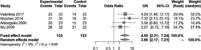
Four articles4–7) could be analyzed for a meta-analysis. Of these, three articles5–7) reported concomitant use of color Doppler. Heterogeneity was not significant, but the overall evidence was described by the random effects. The 3VTV was considered useful for screening (OR, 3.96; 95% CI, 2.17–7.21; p<0.0001).
Three articles1–3) were not included in this meta-analysis. Chen et al1) assessed 77 patients with abnormalities found in the 3VTV in terms of the detection rate of FHD. They reported high sensitivity in the position, the diameter, and the number of the three blood vessels, suggesting that the 3VTV appeared useful. Disappointingly, a control group was not provided in their study. Two meta-analyses2, 3) had some overlap of the original studies referred for the analyses. The results of these meta-analyses suggest that the combined use of three views (the 4CV, the outflow tract view, and the 3VTV) had higher screening sensitivity than the combination of the former two. However, these findings were excluded from this meta-analysis, because the original data could not be confirmed in detail due to descriptions in Chinese or unsearchable references.
No randomized control trial was included in this analysis. The evidence level was C (weak). Even so, the 3VTV is clearly useful as described, with high specificity and low probability of adverse events reported in each study. Therefore, use of the 3VTV is recommended.
Also evaluated was whether the 3VTV was useful for diagnosing certain diseases. Of the four extracted reports,8–11) only one study was included in a meta-analysis to evaluate its usefulness for determining the diagnosis.10) Accordingly, each report is to be discussed.
In a meta-analysis study for diagnosis of coarctation of the aorta (CoA),8) the authors discussed potential predictors using fetal echocardiographic parameters in 72 patients with this malformation. The results suggested that the Z score of the aortic isthmus according to the 3VTV may work as a predictor of the CoA diagnosis. Another study regarding tetralogy of Fallot9) reported that Z score of the aortic isthmus on the 3VTV was likely larger than that of the aortic annulus on the outflow tract view. This result implies that the value obtained on the 3VTV could be used as a marker for diagnosing tetralogy of Fallot. In the remaining two reports,10, 11) the Y-sign and the I-shape sign were found useful when evaluating conotruncal abnormalities. Of 17 patients with tetralogy of Fallot/double outlet right ventricle, 16 (94%) were positive for the Y-sign on the 3VTV. As for complete transposition of the great arteries, the Y-sign was seen in 22 out of 24 patients. The I-shape sign was positive in all these patients. Furthermore, the U-shape or the so-called nine-configuration is a particular finding seen on the 3VTV, representing the right aortic arch or double aortic arch that forms the vascular ring.12, 13) Obviously, the 3VTV is helpful when diagnosing abnormalities of the aortic arch. The vascular diameters and specific features at certain sites obtained on the 3VTV may serve as diagnostic markers. This cross-sectional image is important for diagnosing FHD. Use of the 3VTV is recommended, although the evidence level is C (weak).
Although the strength of evidence is weak, the detection rate seems to be raised by the 3VTV in addition to basic cross-sectional images by the 4CV and the 3VV when screening for CHD. In particular, the vascular diameters at certain sites or specific features on the 3VTV are seemingly useful diagnostic markers. There are few disadvantages of extending the basic cross-sections to the 3VTV, and the potential of adverse events is low. It is highly recommended for the 3VTV to be adopted for screening/diagnosing FHD. The maneuver allows accurate examinations maintaining high levels of sensitivity and specificity.
The 3VTV is used to depict the trachea on the same plane as the pulmonary artery (the ductus arteriosus), the aorta, and the superior vena cava. The 3VTV can be obtained by moving the probe toward the fetal head following the standard fetal ultrasonographic view. The view can be provided relatively easily. Its usefulness at the time of screening and diagnosis of FHD has not yet been clarified. Few studies have been dedicated regarding the role of the 3VTV. In these guidelines, the CQ “Is the 3VTV effective for screening/diagnosing heart disease?” was raised to organize the findings and to make recommendations.
In conclusion, it was suggested that the screening detection rate would be higher by adding the 3VTV in the screening process of the basic cross-sections for FHD. It was also suggested that the diagnosis rate would be higher by using disease-specific markers on the 3VTV when confirming certain diagnoses. This is why use of the 3VTV is highly recommended for screening/diagnosing FHD, despite the weak evidence of its application.
1) Chen KB, Gu Q, Xia T, et al: Three-vessel-trachea view in the diagnosis of fetal cardiac great vessel malformation. J Biol Regul Homeost Agents 2018; 32: 351–355
2) Liu H, Zhou J, Feng QL, et al: Fetal echocardiography for congenital heart disease diagnosis: A meta-analysis, power analysis and missing data analysis. Eur J Prev Cardiol 2015; 22: 1531–1547
3) Zhang YF, Zeng XL, Zhao EF, et al: Diagnostic value of fetal echocardiography for congenital heart disease: A systematic review and meta-analysis. Medicine (Baltimore) 2015; 94: e1759
4) De Robertis V, Rembouskos G, Fanelli T, et al: The three-vessel and trachea view (3VTV) in the first trimester of pregnancy: An additional tool in screening for congenital heart defects (CHD) in an unselected population. Prenat Diagn 2017; 37: 693–698
5) Wiechec M, Knafel A, Nocun A: Prenatal detection of congenital heart defects at the 11- to 13-week scan using a simple color Doppler protocol including the 4-chamber and 3-vessel and trachea views. J Ultrasound Med 2015; 34: 585–594
6) Del Bianco A, Russo S, Lacerenza N, et al: Four chamber view plus three-vessel and trachea view for a complete evaluation of the fetal heart during the second trimester. J Perinat Med 2006; 34: 309–312
7) Wu Q, Li M, Ju L, et al: Application of the 3-vessel view in routine prenatal sonographic screening for congenital heart disease. J Ultrasound Med 2009; 28: 1319–1324
8) Familiari A, Morlando M, Khalil A, et al: Risk factors for coarctation of the aorta on prenatal ultrasound: A systematic review and meta-analysis. Circulation 2017; 135: 772–785
9) Palatnik A, Grobman WA, Cohen LS, et al: Role of the 3-vessel and trachea view in antenatal detection of tetralogy of Fallot. J Ultrasound Med 2016; 35: 1799–1809
10) Pasternok M, Nocun A, Knafel A, et al: “Y Sign” at the level of the 3-vessel and trachea view: An effective fetal marker of aortic dextroposition anomalies in the first trimester. J Ultrasound Med 2018; 37: 1869–1880
11) Palatnik A, Gotteiner NL, Grobman WA, et al: Is the “I-Sign” in the 3-vessel and trachea view a valid tool for prenatal diagnosis of d-transposition of the great arteries? J Ultrasound Med 2015; 34: 1329–1335
12) Razon Y, Berant M, Fogelman R, et al: Prenatal diagnosis and outcome of right aortic arch without significant intracardiac anomaly. J Am Soc Echocardiogr 2014; 27: 1352–1358
13) Bravo C, Gámez F, Pérez R, et al: Fetal aortic arch anomalies: key sonographic views for their differential diagnosis and clinical implications using the cardiovascular system sonographic evaluation protocol. J Ultrasound Med 2016; 35: 237–251
This page was created on 2022-12-26T16:43:48.269+09:00
This page was last modified on 2023-11-27T13:13:17.000+09:00
このサイトは(株)国際文献社によって運用されています。
